. 1998 Oct 5;188(7):1247-54.
doi: 10.1084/jem.188.7.1247.

Regulation of anti-DNA B cells in recombination-activating gene-deficient mice

Affiliations

Regulation of anti-DNA B cells in recombination-activating gene-deficient mice

H Xu et al. J Exp Med. .

Abstract

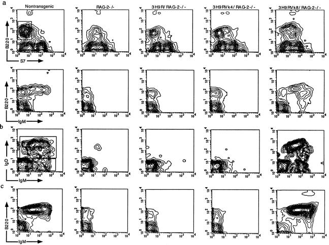
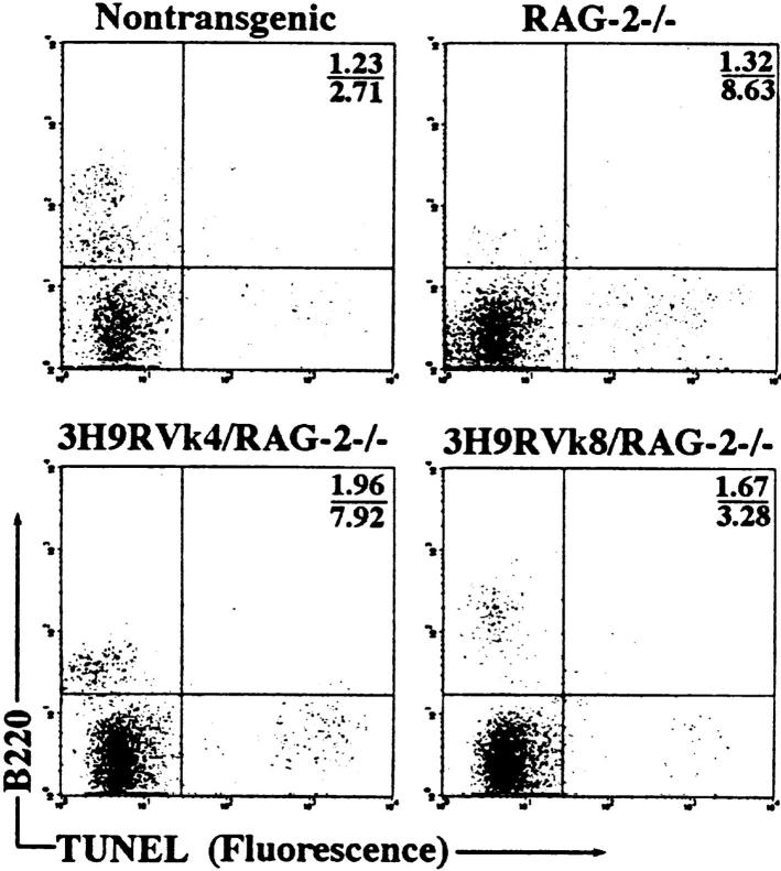
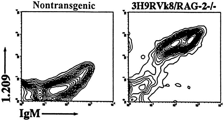
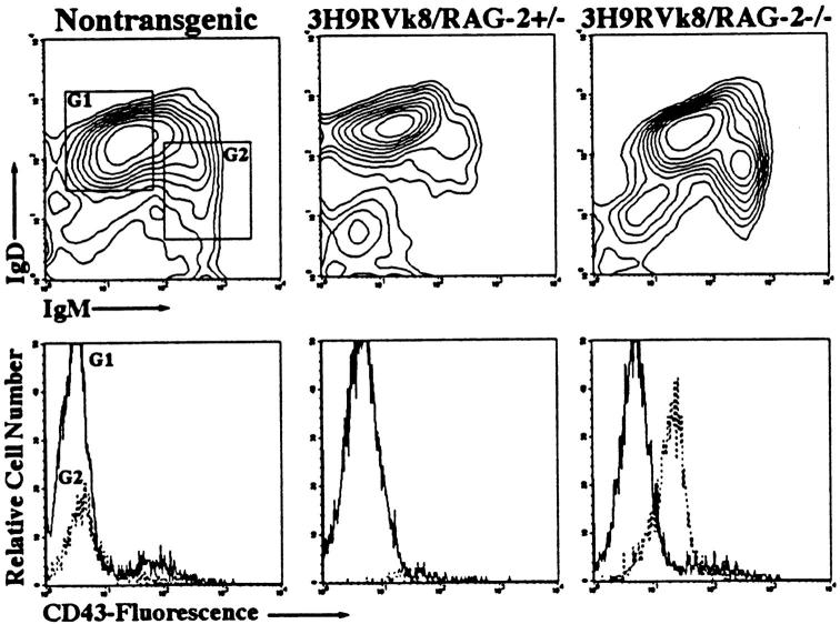
Anti-DNA antibodies are regulated in normal individuals but are found in high concentration in the serum of systemic lupus erythematosus (SLE) patients and the MRL lpr/lpr mouse model of SLE. We previously studied the regulation of anti-double-stranded (ds)DNA and anti-single-stranded (ss)DNA B cells in a nonautoimmune background by generating mice carrying immunoglobulin transgenes coding for anti-DNAs derived from MRL lpr/lpr. Anti-dsDNA B cells undergo receptor editing, but anti-ssDNA B cells seem to be functionally silenced. Here we have investigated how anti-DNA B cells are regulated in recombination- activating gene (RAG)-2-/- mice. In this setting, anti-dsDNA B cells are eliminated by apoptosis in the bone marrow and anti-ssDNA B cells are partially activated.


Figures

Figure 1
Figure 1
Flow cytometric analysis of B cell development. Cells from the bone marrow (a and b) and spleen (c) were resolved by four-color analysis for surface expression of B220/CD45R, CD43/S7, IgM, and IgD. Correlative expression of CD43, B220 (a, top) and IgM (a, bottom) based on lymphocyte gate. Cells from the B220+CD43 window of a (top) were analyzed for expression of IgM and IgD (b). Pre-B (B220+CD43IgM), immature B(B220+CD43IgM+IgD), and mature B (B220+CD43IgM+IgD+) cells are shown in different quadrants. Splenic cells were analyzed with anti-B220 and anti-IgM antibodies based on lymphocyte gate (c).
Figure 2
Figure 2
Analysis of apoptotic cells in the bone marrow by flow cytometry. Bone marrow cells were stained with anti-B220 antibody and fixed with 2% paraformaldehyde. Apoptotic cells were labeled and detected using the In Situ Cell Death Detection Kit (Boehringer Mannheim). The numbers shown represent the percentage of apoptotic cells in the upper right and lower right quadrants.
Figure 3
Figure 3
Binding of 3H9Vk4 antibody to apoptotic cells. (a) DNA fragmentation of apoptotic cells. DNA from freshly isolated splenocytes (lane 1) and cells cultured overnight in the presence of ionomycin (lane 2) was electrophoresed in 1% agar gel. Ionomycin-treated cells show DNA-fragmentation typical of apoptotic cells. (b) Binding of 3H9Vk4 antibody to apoptotic cells. Ionomycin-treated (bold line) and fresh (broken line) spleen cells were incubated with FITC-labeled 3H9Vk4 antibody (left) and the isotype-matched control antibody MOPC141 (right) and analyzed by flow cytometry. (c) Binding of 3H9Vk4 antibody to bone marrow cells. Bone marrow cells of RAG-2−/− (bold line) and control RAG-2+/− (broken line) were isolated, stained with FITC-3H9Vk4 antibody (left) and MOPC 141 (right), and analyzed by flow cytometry.
Figure 3
Figure 3
Binding of 3H9Vk4 antibody to apoptotic cells. (a) DNA fragmentation of apoptotic cells. DNA from freshly isolated splenocytes (lane 1) and cells cultured overnight in the presence of ionomycin (lane 2) was electrophoresed in 1% agar gel. Ionomycin-treated cells show DNA-fragmentation typical of apoptotic cells. (b) Binding of 3H9Vk4 antibody to apoptotic cells. Ionomycin-treated (bold line) and fresh (broken line) spleen cells were incubated with FITC-labeled 3H9Vk4 antibody (left) and the isotype-matched control antibody MOPC141 (right) and analyzed by flow cytometry. (c) Binding of 3H9Vk4 antibody to bone marrow cells. Bone marrow cells of RAG-2−/− (bold line) and control RAG-2+/− (broken line) were isolated, stained with FITC-3H9Vk4 antibody (left) and MOPC 141 (right), and analyzed by flow cytometry.
Figure 4
Figure 4
Staining of B cells with antiidiotype antibody 1.209. Splenocytes were stained with the combinations of anti-IgM and antiidiotype 1.209. Lymphocytes were gated for analysis.
Figure 5
Figure 5
Production of immunoglobulins in 3H9RVk4/RAG-2−/− (○) and 3H9RVk8/RAG-2−/− mice (⋄). Total serum IgM (A) and IgG (B) were determined by ELISA as described in Materials and Methods and compared to normal mice (□).
Figure 5
Figure 5
Production of immunoglobulins in 3H9RVk4/RAG-2−/− (○) and 3H9RVk8/RAG-2−/− mice (⋄). Total serum IgM (A) and IgG (B) were determined by ELISA as described in Materials and Methods and compared to normal mice (□).
Figure 6
Figure 6
FACS® profile of splenocytes from 3H9RVk8/RAG-2−/− mice. Spleen cells were labeled with anti-B220, anti-IgM, anti-IgD, and anti-CD43 antibodies, and gated B220+ cells were analyzed. Cells were then displayed in IgM versus IgD blots and gated for IgMloIgDhi (G1, solid line) and IgMhiIgDlo (G2, broken line) in the upper panel. Either cell population was then analyzed for CD43 expression in the lower panel.
Figure 7
Figure 7
Proliferation of B cells in response to LPS and anti-IgM in absence and presence of IL-4. 2 × 10⁵ T-depleted splenocytes from non-tg (□), 3H9RVk8/RAG-2+/− (▵), and 3H9RVk8/RAG-2−/− (○) mice were cultured in the presence of various doses of LPS (A) and anti-IgM (B) for 72 h. B cells (2 × 10⁵ T cell–depleted splenic cells) were also cultured with submitogenic concentration of LPS (0.1 μg/ml) or anti-IgM (1 μg/ml) in the presence or absence of IL-4 (5 μg/ml) for 72 h (C). Cells were pulsed with [3H]thymidine (1 μCi/well) for 18 h before harvesting. Data shown are the average of triplicate determinations and are representative of at least three different experiments.
Figure 7
Figure 7
Proliferation of B cells in response to LPS and anti-IgM in absence and presence of IL-4. 2 × 10⁵ T-depleted splenocytes from non-tg (□), 3H9RVk8/RAG-2+/− (▵), and 3H9RVk8/RAG-2−/− (○) mice were cultured in the presence of various doses of LPS (A) and anti-IgM (B) for 72 h. B cells (2 × 10⁵ T cell–depleted splenic cells) were also cultured with submitogenic concentration of LPS (0.1 μg/ml) or anti-IgM (1 μg/ml) in the presence or absence of IL-4 (5 μg/ml) for 72 h (C). Cells were pulsed with [3H]thymidine (1 μCi/well) for 18 h before harvesting. Data shown are the average of triplicate determinations and are representative of at least three different experiments.
Figure 8
Figure 8
A model for the regulation of anti-DNA B cells. Pre-B cells that have functional rearranged heavy and light chain genes express surface IgM that can bind to autoantigens. The strong IgM cross-linking by dsDNA from blebs of apoptotic cells induces secondary rearrangement (receptor editing) and altered binding specificity. Anti-dsDNA B cells that fail to edit their receptors (e.g., in the absence of RAG) die via apoptosis. These dying cells provide an enriched source of autoantigens to anti-dsDNA B cells. Weaker IgM cross-linking by ssDNA allows the B cells to mature and populate the periphery with an activated phenotype. We propose that these activated B cells are subsequently inactivated or eliminated by T cell–dependent mechanism(s).

Similar articles

Cited by

References

    1. Tan EM. Antinuclear antibodies: diagnostic markers and clues to the basis of systemic autoimmunity. Pediatr Infect Dis J. 1988;7:S3–S9. - PubMed
    1. Hardin JA. The lupus autoantigens and the pathogenesis of systemic lupus erythematosus. Arthritis Rheum. 1986;29:457–460. - PubMed
    1. Shlomchik MJ, Marshak-Rothstein A, Wolfowicz CB, Rothstein T, Weigert MG. The role of clonal selection and somatic mutation in autoimmunity. Nature. 1987;328:805–811. - PubMed
    1. Shlomchik MJ, Aucoin AH, Pisetsky DS, Weigert MG. Structure and function of anti-DNA autoantibodies derived from a single autoimmune mouse. Proc Natl Acad Sci USA. 1987;84:9150–9154. - PMC - PubMed
    1. Craft JE, Hardin JA. Linked sets of antinuclear antibodies: what do they mean? . J Rheumatol. 1987;14:106–109. - PubMed

Publication types

Substances