. 1998 Oct 5;188(7):1297-306.
doi: 10.1084/jem.188.7.1297.

Different protein tyrosine kinases are required for B cell antigen receptor-mediated activation of extracellular signal-regulated kinase, c-Jun NH2-terminal kinase 1, and p38 mitogen-activated protein kinase

Affiliations

Different protein tyrosine kinases are required for B cell antigen receptor-mediated activation of extracellular signal-regulated kinase, c-Jun NH2-terminal kinase 1, and p38 mitogen-activated protein kinase

A Jiang et al. J Exp Med. .

Abstract

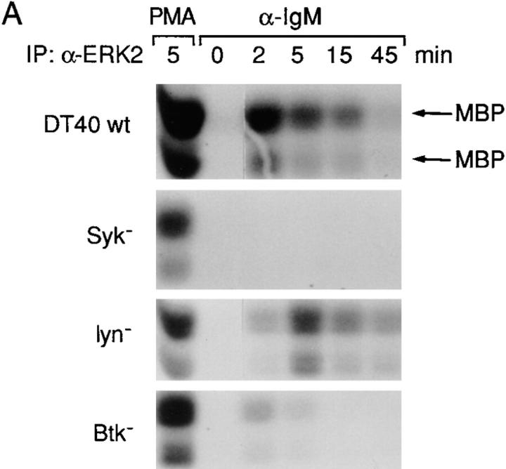
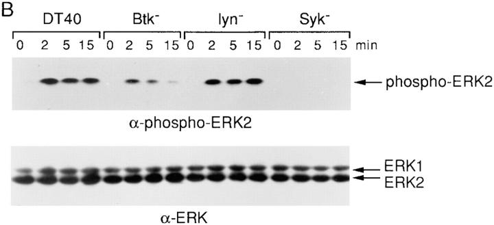
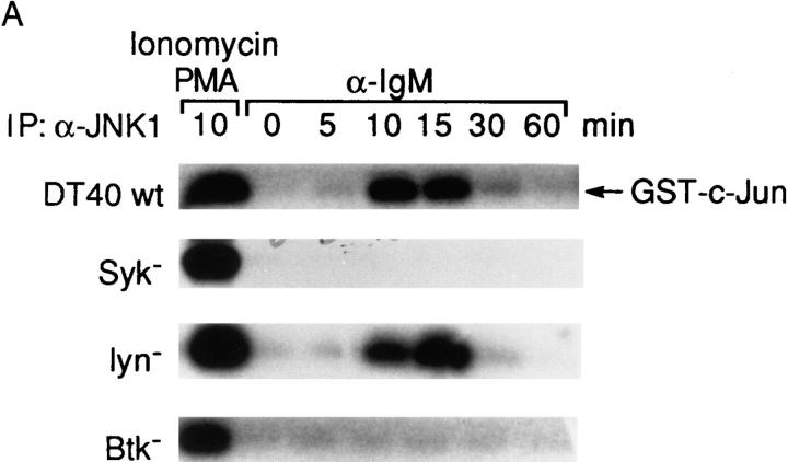
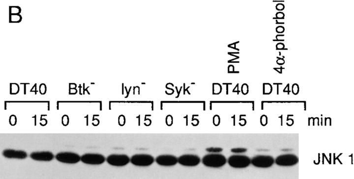
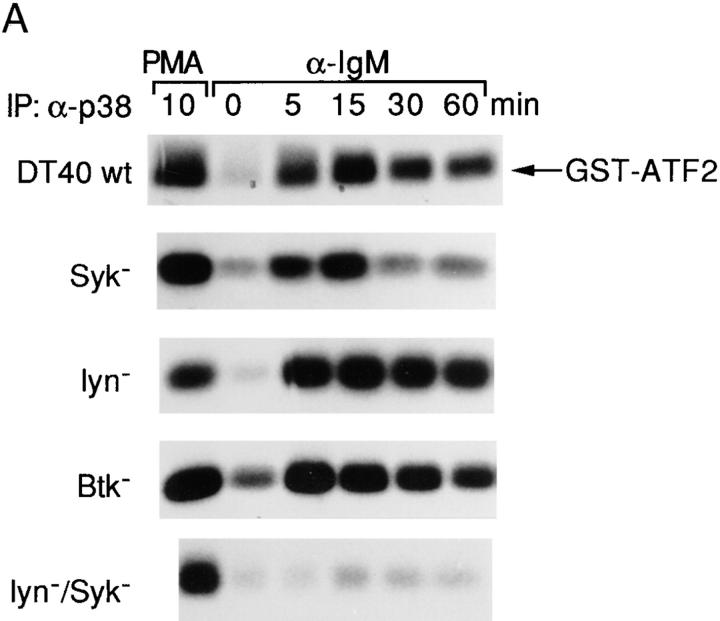
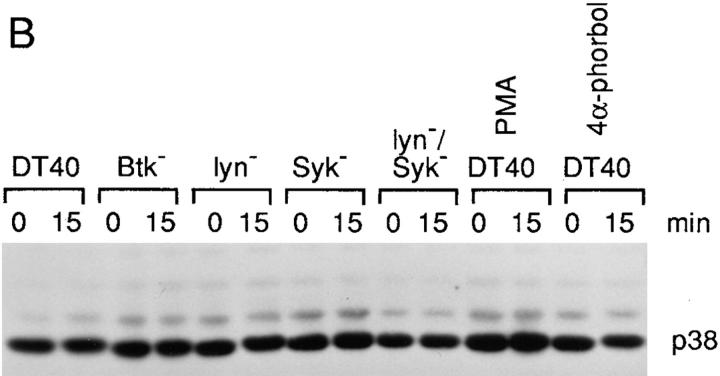
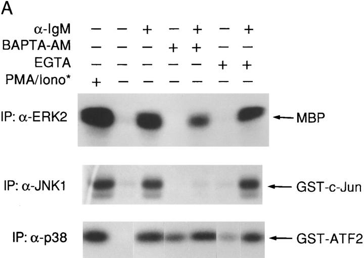
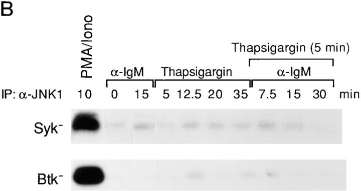
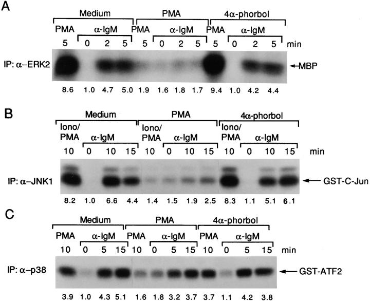
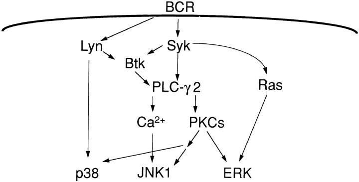
B cell antigen receptor (BCR) cross-linking activates three distinct families of nonreceptor protein tyrosine kinases (PTKs): src-family kinases, Syk, and Btk; these PTKs are responsible for initiating downstream events. BCR cross-linking in the chicken DT40 B cell line also activates three distinct mitogen-activated protein kinases (MAPKs): extracellular signal-regulated kinase (ERK)2, c-jun NH2-terminal kinase (JNK)1, and p38 MAPK. To dissect the functional roles of these PTKs in MAPK signaling, activation of MAPKs was examined in various PTK-deficient DT40 cells. BCR-mediated activation of ERK2, although maintained in Lyn-deficient cells, was abolished in Syk-deficient cells and partially inhibited in Btk-deficient cells, indicating that BCR-mediated ERK2 activation requires Syk and that sustained ERK2 activation requires Btk. BCR-mediated JNK1 activation was maintained in Lyn-deficient cells but abolished in both Syk- and Btk-deficient cells, suggesting that JNK1 is activated via a Syk- and Btk-dependent pathway. Consistent with this, BCR-mediated JNK1 activation was dependent on intracellular calcium and phorbol myristate acetate-sensitive protein kinase Cs. In contrast, BCR-mediated p38 MAPK activation was detected in all three PTK-deficient cells, suggesting that no single PTK is essential. However, BCR-mediated p38 MAPK activation was abolished in Lyn/Syk double deficient cells, demonstrating that either Lyn or Syk alone may be sufficient to activate p38 MAPK. Our data show that BCR-mediated MAPK activation is regulated at the level of the PTKs.


Figures

Figure 1
Figure 1
ERK2 is activated after anti-IgM stimulation in DT40 wild-type, Lyn-, and Btk-deficient cells but not in Syk-deficient cells. (A) Cells (5 × 10⁶ per sample) were stimulated with either anti-IgM mAb M4 (5 μg/ml) for 0–45 min or PMA (50 ng/ ml) for 5 min, and cell lysates were immunoprecipitated with polyclonal anti-ERK2 antiserum (0.5 μg per sample). In vitro kinase assays were performed on the immune complexes using MBP (50 μg/ml) as a substrate; kinase reaction products were resolved by 14% SDS-PAGE, and their activation was quantified by autoradiography and scanning densitometry. MBP appeared as two separated bands under these conditions, reflecting some degradation. One of three similar independent experiments is shown. Note that the ERK2 activation after BCR engagement for 2 min in Lyn-deficient cells was atypically lower than that of DT40 wild-type cells but was similar to that of DT40 wild-type cells in two other experiments. (B) Phosphorylation of ERKs in wild-type and various PTK-deficient DT40 cells. Cells (5 × 10⁶ per sample) were stimulated with anti-IgM mAb M4 (10 μg/ml) for 0–15 min. Cell lysates (equal to 106 per sample) were subsequently analyzed by immunoblotting with anti-phospho-MAPK (top) or anti-ERK1 (bottom) Abs. The anti-ERK1 antibody recognized both ERK1 and ERK2, whereas the anti-phospho-MAPK (ERK2) only detected phosphorylated ERK2 in these DT40 cells. The anti-ERK2 antibody we used for both ERK2 immunoprecipitation and in vitro kinase assays recognized only ERK2 not ERK1 (data not shown).
Figure 1
Figure 1
ERK2 is activated after anti-IgM stimulation in DT40 wild-type, Lyn-, and Btk-deficient cells but not in Syk-deficient cells. (A) Cells (5 × 10⁶ per sample) were stimulated with either anti-IgM mAb M4 (5 μg/ml) for 0–45 min or PMA (50 ng/ ml) for 5 min, and cell lysates were immunoprecipitated with polyclonal anti-ERK2 antiserum (0.5 μg per sample). In vitro kinase assays were performed on the immune complexes using MBP (50 μg/ml) as a substrate; kinase reaction products were resolved by 14% SDS-PAGE, and their activation was quantified by autoradiography and scanning densitometry. MBP appeared as two separated bands under these conditions, reflecting some degradation. One of three similar independent experiments is shown. Note that the ERK2 activation after BCR engagement for 2 min in Lyn-deficient cells was atypically lower than that of DT40 wild-type cells but was similar to that of DT40 wild-type cells in two other experiments. (B) Phosphorylation of ERKs in wild-type and various PTK-deficient DT40 cells. Cells (5 × 10⁶ per sample) were stimulated with anti-IgM mAb M4 (10 μg/ml) for 0–15 min. Cell lysates (equal to 106 per sample) were subsequently analyzed by immunoblotting with anti-phospho-MAPK (top) or anti-ERK1 (bottom) Abs. The anti-ERK1 antibody recognized both ERK1 and ERK2, whereas the anti-phospho-MAPK (ERK2) only detected phosphorylated ERK2 in these DT40 cells. The anti-ERK2 antibody we used for both ERK2 immunoprecipitation and in vitro kinase assays recognized only ERK2 not ERK1 (data not shown).
Figure 2
Figure 2
BCR-induced JNK1 activation requires both Syk and Btk. (A) Activation of JNK1 in wild-type and PTK-deficient DT40 cells. Cells (5 × 10⁶/sample) were stimulated with 5 μg/ ml anti–chicken IgM mAb M4 for 0–60 min or with PMA (50 ng/ml) and ionomycin (250 ng/ ml) for 10 min at 37°C. Cells were then lysed in RIPA buffer and immunoprecipitated with rabbit polyclonal anti-JNK1 antiserum (0.5 μg/sample). In vitro kinase assays were performed with GST–c-jun as a specific exogenous substrate; kinase reaction products were resolved by 12% SDS-PAGE, and their activation was quantified by autoradiography and scanning densitometry. (B) Western blot analysis of JNK1 protein expression. DT40 cells were incubated with PMA (100 ng/ml), the inactive derivative 4α-phorbol (100 ng/ml) or DMSO solvent vehicle control for 24 h before anti-IgM stimulation. Cell lysates (equal to 106/sample) were denatured in SDS sample buffer, resolved by 10% SDS-PAGE, and subsequently were analyzed by Western blot analysis using a mouse anti–human JNK1 monoclonal antibody.
Figure 2
Figure 2
BCR-induced JNK1 activation requires both Syk and Btk. (A) Activation of JNK1 in wild-type and PTK-deficient DT40 cells. Cells (5 × 10⁶/sample) were stimulated with 5 μg/ ml anti–chicken IgM mAb M4 for 0–60 min or with PMA (50 ng/ml) and ionomycin (250 ng/ ml) for 10 min at 37°C. Cells were then lysed in RIPA buffer and immunoprecipitated with rabbit polyclonal anti-JNK1 antiserum (0.5 μg/sample). In vitro kinase assays were performed with GST–c-jun as a specific exogenous substrate; kinase reaction products were resolved by 12% SDS-PAGE, and their activation was quantified by autoradiography and scanning densitometry. (B) Western blot analysis of JNK1 protein expression. DT40 cells were incubated with PMA (100 ng/ml), the inactive derivative 4α-phorbol (100 ng/ml) or DMSO solvent vehicle control for 24 h before anti-IgM stimulation. Cell lysates (equal to 106/sample) were denatured in SDS sample buffer, resolved by 10% SDS-PAGE, and subsequently were analyzed by Western blot analysis using a mouse anti–human JNK1 monoclonal antibody.
Figure 3
Figure 3
Either Lyn or Syk is sufficient for BCR-mediated p38 MAPK activation. (A) BCR-mediated p38 MAPK activation was only abolished in Lyn/syk double deficient cells. Cells (5 × 10⁶ per sample) were stimulated with 10 μg/ml anti–chicken IgM mAb M4 for 0–60 min or with PMA (100 ng/ml) for 10 min at 37°C. Cells were then lysed in RIPA lysis buffer and immunoprecipitated with polyclonal anti-p38 MAPK antiserum (1 μg/sample) in the presence of an additional 0.1% SDS. In vitro kinase assays were performed using GST-ATF2 as a specific exogenous substrate and analyzed as described in the legend to Fig. 2 A. (B) Western blot analysis of p38 MAPK expression. Cell lysates were prepared as described in the legend to Fig. 2 B. The lysates (equal to 106/ sample) were resolved by 10% SDS-PAGE and subsequently were analyzed by Western blot analysis using a polyclonal anti-p38 MAPK antiserum.
Figure 3
Figure 3
Either Lyn or Syk is sufficient for BCR-mediated p38 MAPK activation. (A) BCR-mediated p38 MAPK activation was only abolished in Lyn/syk double deficient cells. Cells (5 × 10⁶ per sample) were stimulated with 10 μg/ml anti–chicken IgM mAb M4 for 0–60 min or with PMA (100 ng/ml) for 10 min at 37°C. Cells were then lysed in RIPA lysis buffer and immunoprecipitated with polyclonal anti-p38 MAPK antiserum (1 μg/sample) in the presence of an additional 0.1% SDS. In vitro kinase assays were performed using GST-ATF2 as a specific exogenous substrate and analyzed as described in the legend to Fig. 2 A. (B) Western blot analysis of p38 MAPK expression. Cell lysates were prepared as described in the legend to Fig. 2 B. The lysates (equal to 106/ sample) were resolved by 10% SDS-PAGE and subsequently were analyzed by Western blot analysis using a polyclonal anti-p38 MAPK antiserum.
Figure 4
Figure 4
BCR-mediated ERK2, JNK1, and p38 MAPK activation have different requirements of calcium. (A) Anti-IgM–induced activation of JNK1, but not of ERK2 and p38 MAPK, is dependent on intracellular calcium. Cells were preincubated with BAPTA-AM (10 μM) for 10 min or EGTA (2 μM) for 30 min and then stimulated with anti-IgM mAb M4 (10 μg/ml). The concentrations of BAPTA-AM and EGTA were titrated to prevent calcium release or extracellular calcium influx, respectively (data not shown). In vitro kinase assays were performed to measure the activation of ERK2, JNK1 and p38 MAPK as described in the legends to Figs. 1–3, except that 12% SDS-PAGE was used for all these assays. The times of stimulation of cells were as follows: ERK2, PMA for 5 min or anti-IgM for 2 min; JNK1, PMA and ionomycin or anti-IgM for 10 min; and p38 MAPK, PMA for 10 min or anti-IgM for 15 min. (B) A calcium signal is not sufficient for anti-IgM–induced JNK1 activation. Btk- and Syk-deficient cells were pre-incubated with thapsigargin (10 nM) for 5 min and then stimulated with anti-IgM mAb M4 (10 μg/ml) or incubated with thapsigargin alone for the indicated times. JNK1 activation was quantified by in vitro kinase assays as described in the legend to Fig. 2 A.
Figure 4
Figure 4
BCR-mediated ERK2, JNK1, and p38 MAPK activation have different requirements of calcium. (A) Anti-IgM–induced activation of JNK1, but not of ERK2 and p38 MAPK, is dependent on intracellular calcium. Cells were preincubated with BAPTA-AM (10 μM) for 10 min or EGTA (2 μM) for 30 min and then stimulated with anti-IgM mAb M4 (10 μg/ml). The concentrations of BAPTA-AM and EGTA were titrated to prevent calcium release or extracellular calcium influx, respectively (data not shown). In vitro kinase assays were performed to measure the activation of ERK2, JNK1 and p38 MAPK as described in the legends to Figs. 1–3, except that 12% SDS-PAGE was used for all these assays. The times of stimulation of cells were as follows: ERK2, PMA for 5 min or anti-IgM for 2 min; JNK1, PMA and ionomycin or anti-IgM for 10 min; and p38 MAPK, PMA for 10 min or anti-IgM for 15 min. (B) A calcium signal is not sufficient for anti-IgM–induced JNK1 activation. Btk- and Syk-deficient cells were pre-incubated with thapsigargin (10 nM) for 5 min and then stimulated with anti-IgM mAb M4 (10 μg/ml) or incubated with thapsigargin alone for the indicated times. JNK1 activation was quantified by in vitro kinase assays as described in the legend to Fig. 2 A.
Figure 5
Figure 5
ERK2, JNK1, and p38 MAPK have different requirements for PMA- depletable PKCs. Cells (5 × 10⁶/sample) were incubated with PMA (100 ng/ml), the inactive derivative 4α-phorbol (100 ng/ml), or DMSO solvent vehicle control for 24 h before anti-IgM stimulation. Cell lysates (5 × 10⁶/sample) were then prepared, and ERK2, JNK1, and p38 MAPK activation was measured as described in the legends to Figs. 1–3, except that 12% SDS-PAGE was used in all cases.
Figure 6
Figure 6
A model for the different pathways leading to BCR-mediated ERK2, JNK1, and p38 MAPK activation in DT40 cells. BCR cross-linking leads to activation of the protein tyrosine kinases Lyn, Syk, and Btk. Loss of either Syk or Btk leads to abrogation of calcium mobilization, although only Syk is essential for PLC-γ2 phosphorylation. PLC-γ2 phosphorylation leads to calcium mobilization and PKC activation. Syk but not Lyn is essential for BCR-mediated ERK2 activation and Btk may function as a regulator of the ERK2 pathway since partial BCR-mediated ERK2 activation is observed in Btk-deficient cells. The requirement for Syk and Btk may reflect the requirement of PKCs for ERK2 activation. In contrast, BCR-mediated JNK1 activation depends on both Syk and Btk but not Lyn, via a calcium- and PKC-dependent pathway. For p38 MAPK, a distinct pathway is used for BCR-mediated activation of p38 MAPK, since either Syk or Lyn is sufficient for BCR-mediated p38 MAPK activation. A Lyn-dependent pathway for p38 MAPK activation may not require PKCs, whereas the Syk-dependent pathway does. Neither a calcium signal nor PMA-depletable PKCs are absolutely essential for BCR-mediated p38 MAPK activation, although PMA-insensitive PKCs may be required.

Similar articles

Cited by

References

    1. Campbell KS, Cambier JC. B lymphocyte antigen receptors (mIg) are non-covalently associated with a disulfide linked, inducibly phosphorylated glycoprotein complex. EMBO (Eur Mol Biol Organ) J. 1990;9:441–448. - PMC - PubMed
    1. Reth M. Antigen receptors on B lymphocytes. Annu Rev Immunol. 1992;10:97–121. - PubMed
    1. Bolen JB. Protein tyrosine kinases in the initiation of antigen receptor signaling. Curr Opin Immunol. 1990;7:306–311. - PubMed
    1. Sefton BM, Campbell MA. The role of tyrosine protein phosphorylation in lymphocyte activation. Annu Rev Cell Biol. 1991;7:257–274. - PubMed
    1. Law CL, Craxton A, Otipoby KL, Sidorenko SP, Klaus SJ, Clark EA. Regulation of signalling through B-lymphocyte antigen receptors by cell–cell interaction molecules. Immunol Rev. 1996;153:123–154. - PubMed

Publication types

MeSH terms

Substances