. 1998 Oct 5;188(7):1307-20.
doi: 10.1084/jem.188.7.1307.

Expression of interleukin 9 in the lungs of transgenic mice causes airway inflammation, mast cell hyperplasia, and bronchial hyperresponsiveness

Affiliations

Expression of interleukin 9 in the lungs of transgenic mice causes airway inflammation, mast cell hyperplasia, and bronchial hyperresponsiveness

U A Temann et al. J Exp Med. .

Abstract

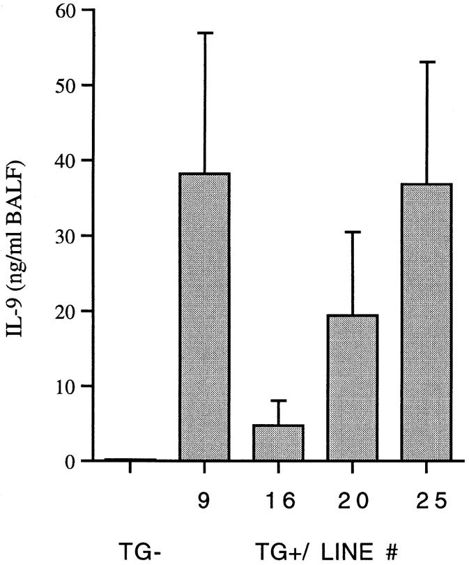
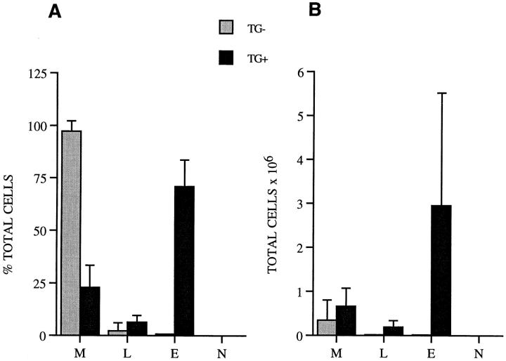
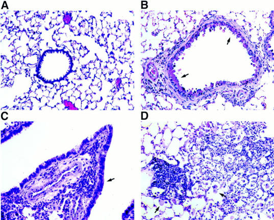
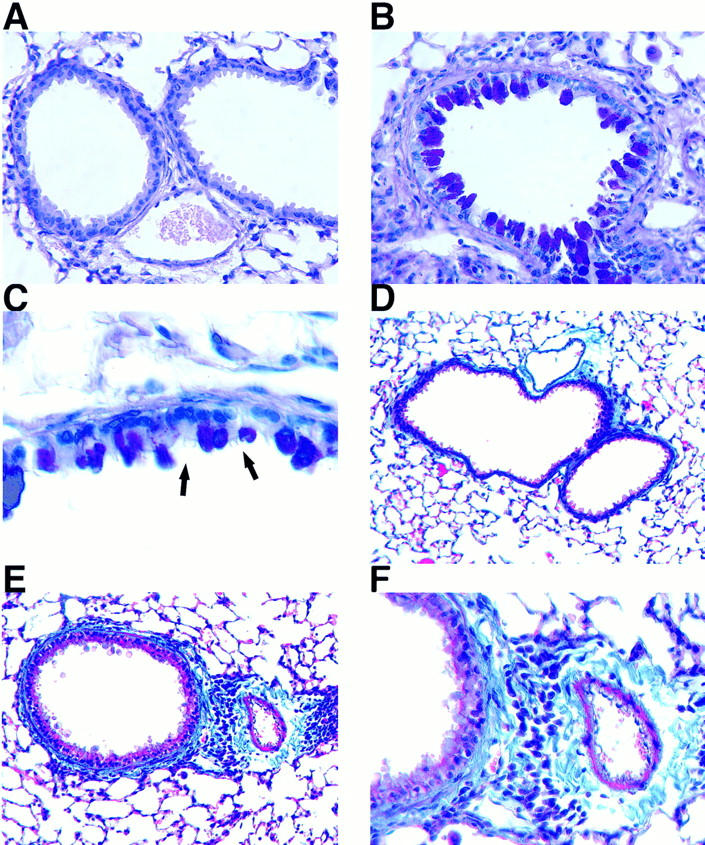
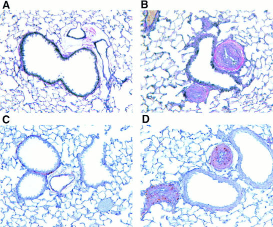
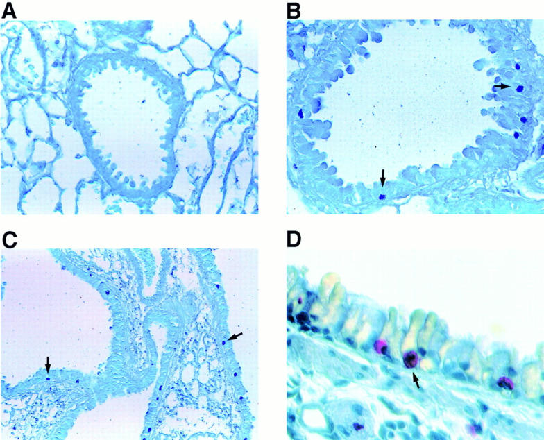
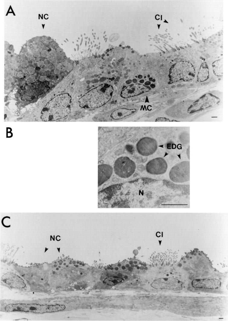
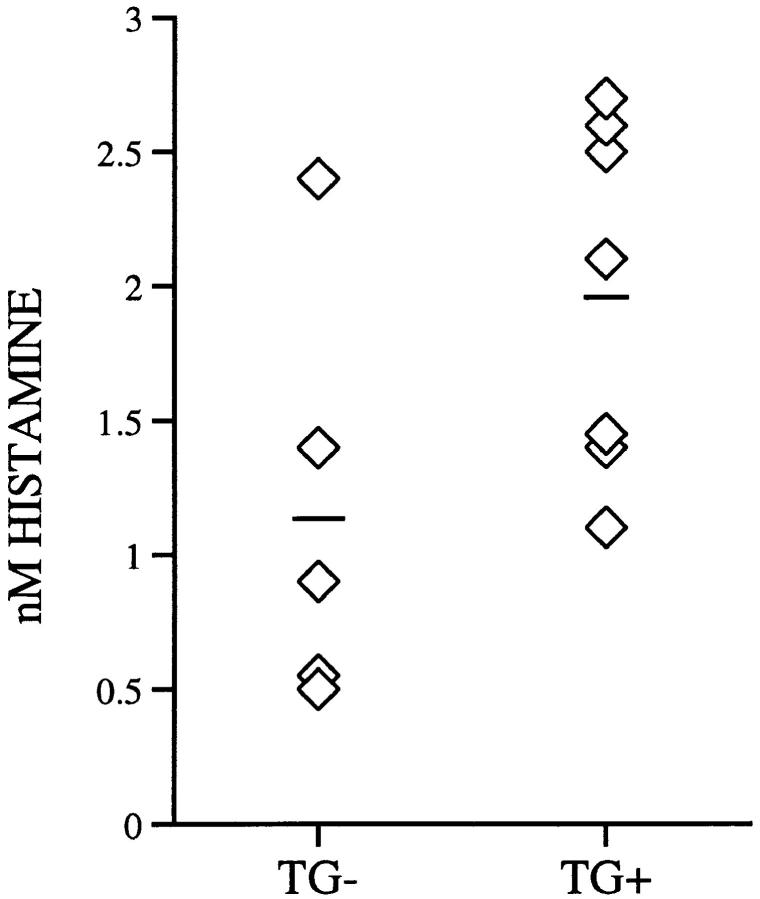
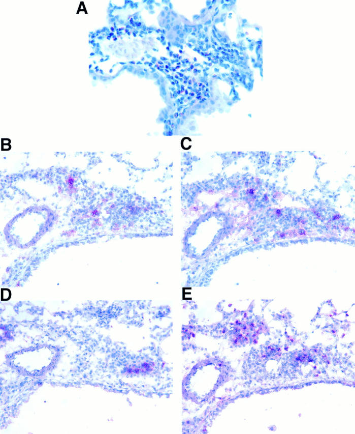
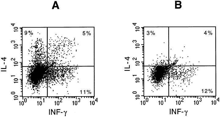
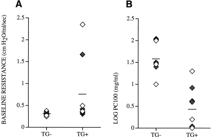
Interleukin (IL)-9, a pleiotropic cytokine produced by the Th2 subset of T lymphocytes has been proposed as product of a candidate gene responsible for asthma. Its wide range of biological functions on many cell types involved in the allergic immune response suggests a potentially important role in the complex pathogenesis of asthma. To investigate the contributions of IL-9 to airway inflammation and airway hyperresponsiveness in vivo, we created transgenic mice in which expression of the murine IL-9 cDNA was regulated by the rat Clara cell 10 protein promoter. Lung selective expression of IL-9 caused massive airway inflammation with eosinophils and lymphocytes as predominant infiltrating cell types. A striking finding was the presence of increased numbers of mast cells within the airway epithelium of IL-9-expressing mice. Other impressive pathologic changes in the airways were epithelial cell hypertrophy associated with accumulation of mucus-like material within nonciliated cells and increased subepithelial deposition of collagen. Physiologic evaluation of IL-9-expressing mice demonstrated normal baseline airway resistance and markedly increased airway hyperresponsiveness to inhaled methacholine. These findings strongly support an important role for IL-9 in the pathogenesis of asthma.


Figures

Figure 1
Figure 1
IL-9 levels in bronchoalveolar lavage fluid (BALF ). Transgene-positive animals (TG+) from four independent lines (9, 16, 20, and 25) expressed different amounts of IL-9. Each column represents the mean ± SD of three representative animals. No IL-9 was detectable in lung lavage fluid from transgene-negative animals (TG−).
Figure 2
Figure 2
Inflammatory cells in lung lavage fluid. Differential cell counts were derived from at least 500 cell counts and are presented as percentage of each cell type from total cells (A) and as total cell numbers (B). Data are the mean ± SD of counts from 13 IL-9–expressing mice (black bars) and four transgene-negative control mice (gray bars) from all four transgenic lines. The number of eosinophils (E) and lymphocytes (L) were significantly increased in IL-9–expressing mice compared with transgene-negative mice (P ≤ 0.04 and P < 0.03; B), whereas the number of macrophages (M) was only slightly but not significantly increased (B). N, neutrophils.
Figure 3
Figure 3
Lung histology of conducting airways and parenchyma. Sections of formalin-fixed lung tissue from a transgene-negative control mouse (A) and an IL-9–expressing mouse of line 9 (B–D) were stained with hematoxylin and eosin before examination by light microscopy. Lung sections from IL-9–expressing mice revealed the presence of inflammatory cells in the subepithelium of conducting airways of all different sizes, smaller airways (B) and larger airways (C), and around blood vessels, which was not seen in sections from transgene-negative mice (A). The airway epithelium was hypertrophic (B and C, arrows) compared with that in transgene-negative animals (A). Cellular infiltrations in the lung parenchyma from IL-9–expressing mice were often associated with nodule-like accumulations of mononuclear cells and large macrophages (D, arrows). Original magnification, A–D: ×250.
Figure 4
Figure 4
Histologic staining for mucin and collagen in lung sections. Light micrographs of formalin-fixed tissue from a transgene-negative control mouse (A and D) and an IL-9–expressing mouse (B, C, E, and F ) stained with alcian blue/periodic acid-Schiff (A–C) or Masson's trichrome (D–F ). The hypertrophic airway epithelium in sections from IL-9–expressing mice stained positively (magenta) for mucins with alcian blue/periodic acid–Schiff (B). Higher magnification of the epithelium revealed that only nonciliated epithelial cells but not ciliated cells (arrows) were hypertrophic and stained positive for mucin (C). The airway epithelium in sections from transgene–negative mice did not stain for mucin (A). Lung sections stained with Masson's trichrome demonstrated that thickening of the airway wall in IL-9–expressing mice is associated with increased deposition of material staining positively (blue) for collagen (E), which was not present in transgene-negative mice (D). A higher magnification of the same area demonstrates extracellular, intensely blue staining collagen adjacent to inflammatory cells around airway and blood vessel (F ). Original magnifications: A, B, and F, ×500; C, ×1,000; D and E, ×250.
Figure 5
Figure 5
Histologic analysis of blood vessels. Blood vessels in van Gieson–stained sections from formalin-fixed lung tissue of an IL-9–expressing mouse (B) showed medial hypertrophy compared with vessels from a transgene-negative control mouse (A). Immunostaining for α-smooth muscle actin was strongly positive (brown) in wall and lumen of blood vessels found in sections from an IL-9–expressing mouse (D), which was not observed in blood vessels from transgene-negative mice (C). Original magnification, A–D, ×500.
Figure 6
Figure 6
Histologic staining for mast cells. Toluidine blue staining of sections from formalin-fixed lung tissue revealed mast cells in the airway epithelium from an IL-9–expressing mouse (B and C) but not in the epithelium from transgene-negative mice (A). Dark blue–stained mast cell granules (arrows) were present in the epithelium of smaller (B) and larger (C) airways. Higher magnification shows red-stained mast cells (arrow) in the airway epithelium using Leder's chloracetate esterase reaction (D). Original magnifications: A and B, ×500; C, ×250; D, ×1,000.
Figure 7
Figure 7
Electron micrographs of epithelium from conducting airways of the lower respiratory tract. Representative area of the airway epithelium from an IL-9–expressing mouse shows that fully granulated mast cells (MC) were present (A). A higher magnification of mast cell granules from the same cell as shown in A did not reveal any signs of degranulation (B). N, nucleus, EDG, electron-dense granules. Mast cells were not present in airway epithelium from transgene-negative control mice (C). Nonciliated epithelial cells (NC) in IL-9–expressing mice were hypertrophic and showed accumulations of low-density vacuoles, whereas ciliated cells (CI ) appeared normal (A). Epithelium from a transgene-negative mouse shows normal nonciliated (NC) and ciliated (CI ) epithelial cells (C). Bars in A, B, and C represent 1 μm. Original magnifications: A, ×5,000; B, ×22,400; C, ×3,300.
Figure 8
Figure 8
Histamine levels in lung lavage fluid. IL-9–expressing mice (TG+) and transgene negative mice (TG−) had similar histamine levels in lung lavage fluid as determined by enzyme immunoassay. Levels are expressed as individual data points for each mouse (diamonds) and as means (lines).
Figure 9
Figure 9
Characterization of inflammatory cells in lung sections from an IL-9–expressing mouse. Formalin-fixed tissue was stained with new vital red to demonstrate that eosinophils were present in high numbers in lung tissue from IL-9–expressing mice (A). Immunostaining of frozen lung sections (B–E) revealed that cell infiltrates also contained CD4+ (B), CD8+ (C), B220+ (D), and Mac-1+ (E) mononuclear cells. Original magnifications: A, ×600; B–E, ×500.
Figure 10
Figure 10
Intracellular detection of IL-4 and INF-γ in lymphocytes from lung lavage fluid of IL-9–expressing mice. Data shown are generated by three-color flow cytometric analysis of total lung lavage cells and pooled from four IL-9–expressing littermates after short-term culture in the presence of PMA, ionomycin, and monensin. Dot-blots were gated on CD4+ lymphocytes (A) or CD8+ lymphocytes (B). Number of cells staining for each cytokine are expressed as a percentage of CD4+ or CD8+ cells.
Figure 11
Figure 11
Assessment of lung physiology. Two independent experiments were performed to compare airway baseline resistance (A) and airway hyperresponsiveness to inhaled methacholine, LOG PC100 (B), in IL-9–expressing mice (TG+) and in transgene-negative littermates (TG−). Mice derived from two independent lines, 9 (open diamonds) and 25 ( filled diamonds), are expressed as individual data points and as means (lines).

References

    1. Kay AB. Asthma and inflammation. J Allergy Clin Immunol. 1991;87:893–910. - PubMed
    1. Lukacs NW, Strieter RM, Kunkel SL. Leukocyte infiltration in allergic airway inflammation. Am J Respir Cell Mol Biol. 1995;13:1–6. - PubMed
    1. Busse WW, Coffman RL, Gelfand EW, Kay AB, Rosenwasser LJ. Mechanisms of persistant airway inflammation. Am J Respir Crit Care Med. 1995;152:388–393. - PubMed
    1. Barnes PJ. Cytokines as mediators of chronic asthma. Am J Respir Crit Care Med. 1994;150:S42–S49. - PubMed
    1. Lukacs NW, Strieter RM, Chensue SW, Kunkel SL. Activation and regulation of chemokines in allergic airway inflammation. J Leukocyte Biol. 1996;59:13–17. - PubMed

Publication types

MeSH terms