. 1998 Oct 5;188(7):1369-73.
doi: 10.1084/jem.188.7.1369.

The interleukin 2 receptor alpha chain/CD25 promoter is a target for nuclear factor of activated T cells

Affiliations

The interleukin 2 receptor alpha chain/CD25 promoter is a target for nuclear factor of activated T cells

K Schuh et al. J Exp Med. .

Abstract

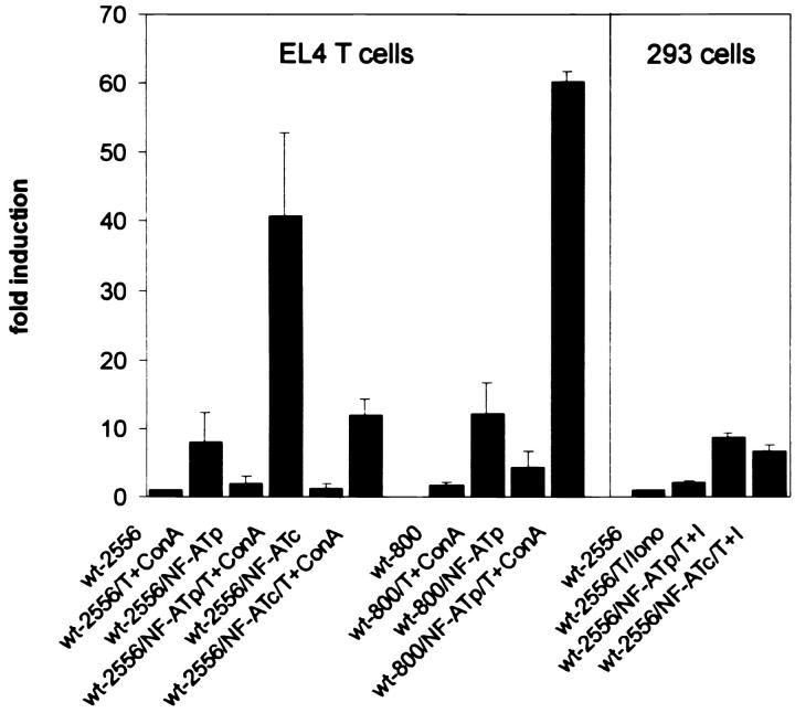
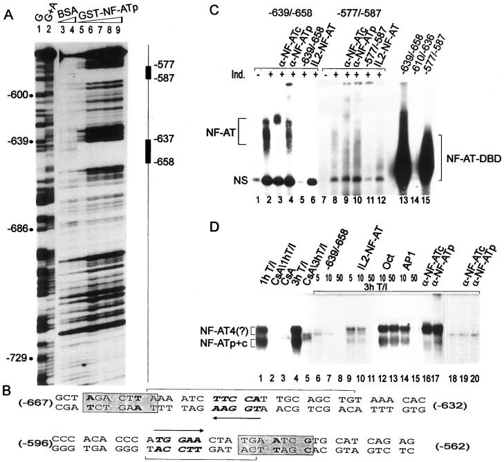
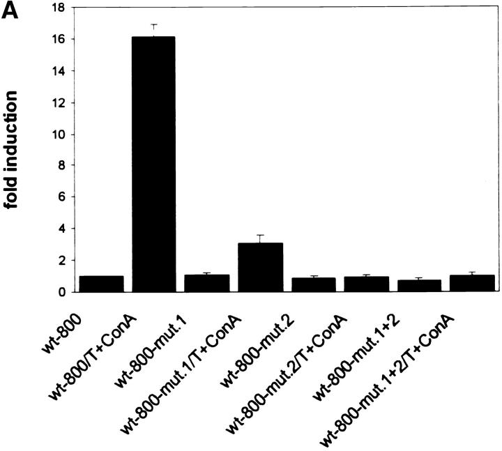
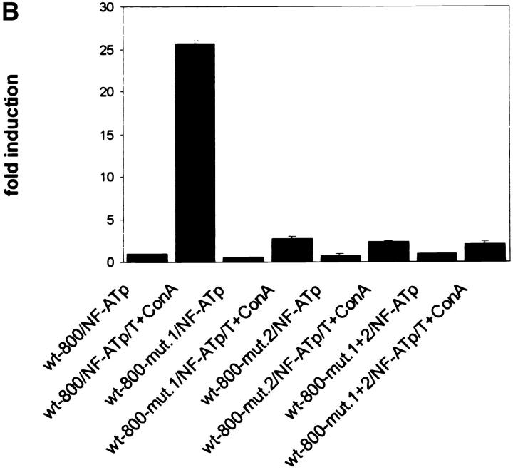
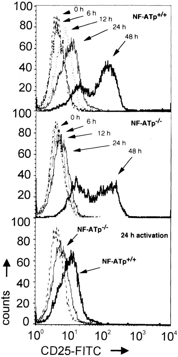
The expression of the murine interleukin (IL)-2 receptor alpha chain/CD25 is strongly induced at the transcriptional level after T cell activation. We show here that nuclear factor of activated T cell (NF-AT) factors are involved in the control of CD25 promoter induction in T cells. NF-ATp and NF-ATc bind to two sites around positions -585 and -650 located upstream of the proximal CD25 promoter. Immediately 3' from these NF-AT motifs, nonconsensus sites are located for the binding of AP-1-like factors. Mutations of sites that suppress NF-AT binding impair the induction and strong NF-ATp-mediated transactivation of the CD25 promoter in T cells. In T lymphocytes from NF-ATp-deficient mice, the expression of CD25 is severely impaired, leading to a delayed IL-2 receptor expression after T cell receptor (TCR)/CD3 stimulation. Our data indicate an important role for NF-AT in the faithful expression of high affinity IL-2 receptors and a close link between the TCR-mediated induction of IL-2 and IL-2 receptor alpha chain promoters, both of which are regulated by NF-AT factors.


Figures

Figure 1
Figure 1
NF-ATp transactivates the murine CD25 promoter in T cells. 2.5 μg DNA of luciferase reporter gene constructs controlled by murine CD25 promoters up to position −2556 (wt-2556) and −800 (wt-800) were transfected into murine El4 T thymoma cells or human embryonic 293 kidney cells, along with an empty RSV-based expression vector or vectors expressing NF-ATp (10) or NF-ATc. The cells were induced as indicated for 18 h. To calculate the extent of induction, the activity of the CD25 wild-type promoter in nonstimulated cells was used as a reference point (onefold).
Figure 2
Figure 2
Binding of NF-AT to the CD25 promoter. (A) DNase I footprint protection assay. A CD25 promoter probe spanning the nucleotides from −548 to −804 was incubated with 3 and 5 μg BSA (lanes 3 and 4) or 1–5 μg GST–NF-ATp protein (lanes 5–9). The NF-ATp–specific footprints are indicated. G and G+A, chemical sequencing reactions. (B) Sequences of footprint regions. The footprints are indicated in brackets. The arrows indicate the direction of TGGAA NF-AT “core” motifs. The TPA responsive element–like sequence motifs 3′ from the NF-AT motifs are boxed. (C) EMSAs with the NF-AT sites. In lanes 1–12, nuclear proteins from murine splenocytes are shown, and in lanes 13–15, GST–NF-ATp was used with the −639/−658 (lanes 1–6 and 13) and −577/−587 probes (lanes 7–12 and 15). In lane 14, a probe of the −610/−636 site was used as a control. 2 μg of nuclear proteins from uninduced splenocytes (−) or splenocytes induced for 2 h with T+I (+) were incubated in the absence or presence of 0.5 μg NF-ATc– or NF-ATp–specific Abs, or in the presence of a 200-fold molar excess of homologous unlabeled probes or distal IL-2 NF-AT site as indicated. Note that the autoradiograph of lanes 1–6 was exposed for 16 h, and that of lanes 7–12 for 3 d. NS, nonspecific complex. Free probes are cut off. (D) EMSAs of the −639/−658 site using nuclear proteins from Jurkat cells. A probe of the −639/−658 site was incubated with 2 μg of nuclear proteins from Jurkat cells treated with T+I in the absence or presence of 100 ng/ml cyclosporin A (CsA). For competition the following DNAs were used: 5–50 ng of the cold −639/−658 site and the distal IL-2 NF-AT site, or 10 and 50 ng of the proximal IL-2 octamer site (11) and a consensus AP-1 site. In lanes 16 and 17, 0.5 μg of NF-AT–specific Abs were added. In lanes 18–20, a −632/−667 probe mutated in the NF-AT site (see oligonucleotide a in Materials and Methods) was incubated without or with NF-AT–specific Abs.
Figure 3
Figure 3
The NF-ATp sites contribute to the induction of CD25 promoter. (A) Mutations within the NF-ATp sites that suppress NF-AT binding interfere with the T+Con A–mediated CD25 promoter induction in El4 cells. 2.5 μg of luciferase constructs containing the wild-type CD25 promoter up to −800 bp or a promoter with mutations in the −658/−639 (mut. 1), the −587/−577 site (mut. 2), or in both sites (mut. 1+2) was transfected into El4 cells that were induced for 12 h. (B) Mutations of NF-AT sites suppress the NF-ATp–mediated transactivation of the CD25 promoter. 0.5 μg of the CD25 luciferase constructs was cotransfected with an NF-ATp expression vector into El4 cells that were stimulated for 12 h. To calculate the extent of induction, the activity of the CD25 wild-type promoter in nonstimulated cells was used as a reference point (onefold).
Figure 3
Figure 3
The NF-ATp sites contribute to the induction of CD25 promoter. (A) Mutations within the NF-ATp sites that suppress NF-AT binding interfere with the T+Con A–mediated CD25 promoter induction in El4 cells. 2.5 μg of luciferase constructs containing the wild-type CD25 promoter up to −800 bp or a promoter with mutations in the −658/−639 (mut. 1), the −587/−577 site (mut. 2), or in both sites (mut. 1+2) was transfected into El4 cells that were induced for 12 h. (B) Mutations of NF-AT sites suppress the NF-ATp–mediated transactivation of the CD25 promoter. 0.5 μg of the CD25 luciferase constructs was cotransfected with an NF-ATp expression vector into El4 cells that were stimulated for 12 h. To calculate the extent of induction, the activity of the CD25 wild-type promoter in nonstimulated cells was used as a reference point (onefold).
Figure 4
Figure 4
Impaired CD25 expression on NF-ATp−/− LN T cells. LN T cells from NF-ATp+/+ and NF-ATp−/− mice (13) were treated with plate-bound α-CD3 mAb in vitro. The cells were stained with mAbs directed against murine CD25 (7D4; PharMingen) and α/β-TCR (H57-597; PharMingen). The expression of CD25 on α/β-TCR+ LN cells is shown. The lowest panel shows a comparison of CD25 expression on NF-ATp+/+ and NF-ATp−/− TCR+ LN cells stimulated for 24 h. The dotted line indicates the isotype control staining of LN cells.

References

    1. Minami J, Kono T, Miyazaki T, Taniguchi T. The IL-2 receptor complex: its structure, function, and target genes. Annu Rev Immunol. 1993;11:245–268. - PubMed
    1. Sperisen P, Wang SM, Soldaini E, Pla M, Rusterholz C, Bucher P, Corthesy P, Reichenbach P, Nabholz M. Mouse interleukin-2 receptor α gene expression. Interleukin-1 and interleukin-2 control transcription via distinct cis-acting elements. J Biol Chem. 1995;277:10743–10752. - PubMed
    1. John S, Reeves RB, Lin J-Y, Child R, Leiden JM, Thompson CB, Leonard WJ. Regulation of cell-type–specific interleukin-2 receptor α chain gene expression: potential role of physical interactions between Elf-1, HMG-I(Y), and NF-κB family proteins. Mol Cell Biol. 1995;15:1786–1796. - PMC - PubMed
    1. Serdobova I, Pla M, Reichenbach P, Sperisen P, Ghysdael J, Wilson A, Freeman J, Nabholz M. Elf-1 contributes to the function of the complex interleukin (IL)- 2–responsive enhancer in mouse IL-2 receptor α gene. J Exp Med. 1997;185:1211–1221. - PMC - PubMed
    1. John S, Robbins CM, Leonard WJ. An IL-2 response element in the human IL-2 receptor α chain promoter is a composite element that binds Stat5, Elf-1, HMG-I(Y) and a GATA family protein. EMBO (Eur Mol Biol Organ) J. 1996;15:5627–5635. - PMC - PubMed

Publication types