. 1998 Oct 5;188(7):1385-90.
doi: 10.1084/jem.188.7.1385.

Regulation of L-selectin-mediated rolling through receptor dimerization

Affiliations

Regulation of L-selectin-mediated rolling through receptor dimerization

X Li et al. J Exp Med. .

Abstract

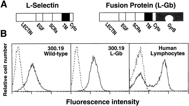
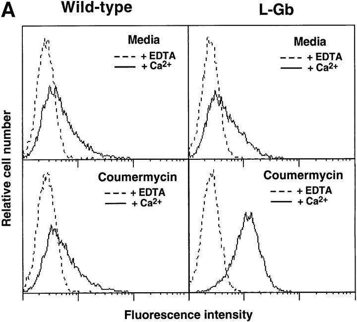
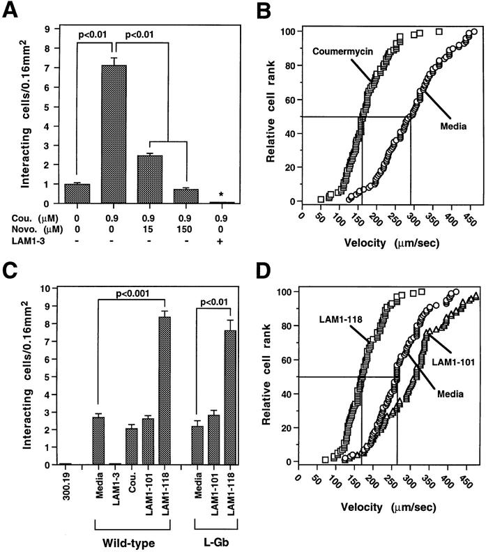
L-selectin binding activity for its ligand expressed by vascular endothelium is rapidly and transiently increased after leukocyte activation. To identify mechanisms for upregulation and assess how this influences leukocyte/endothelial cell interactions, cell-surface dimers of L-selectin were induced using the coumermycin-GyrB dimerization strategy for cross-linking L-selectin cytoplasmic domains in L-selectin cDNA-transfected lymphoblastoid cells. Coumermycin- induced L-selectin dimerization resulted in an approximately fourfold increase in binding of phosphomanan monoester core complex (PPME), a natural mimic of an L-selectin ligand, comparable to that observed after leukocyte activation. Moreover, L-selectin dimerization significantly increased (by approximately 700%) the number of lymphocytes rolling on vascular endothelium under a broad range of physiological shear stresses, and significantly slowed their rolling velocities. Therefore, L-selectin dimerization may explain the rapid increase in ligand binding activity that occurs after leukocyte activation and may directly influence leukocyte migration to peripheral lymphoid tissues or to sites of inflammation. Inducible oligomerization may also be a common mechanism for rapidly upregulating the adhesive or ligand-binding function of other cell-surface receptors.


Figures

Figure 1
Figure 1
Generation of L-selectin–expressing cell lines. (A) Structure of L-selectin and the L-selectin–GyrB (L-Gb) fusion protein containing the entire L-selectin protein in-frame with the NH2-terminal 24-kD subdomain of the B subunit of bacterial DNA gyrase (GyrB). Domains: EGF, epidermal growth factor–like; SCR, short consensus repeat; TM, transmembrane; Cyto, cytoplasmic. (B) Cell-surface expression of wild-type L-selectin or L-Gb in stably-transfected 300.19 cells and wild-type L-selectin expression by human blood lymphocytes. Cells were isolated and stained with FITC-conjugated LAM1-116 mAb specific for L-selectin (solid line) or an isotype-matched, nonbinding control mAb (dashed line) as previously described (16). Fluorescence histograms from flow cytometry analysis are on a three-decade log scale and are representative of results from at least five experiments.
Figure 2
Figure 2
Coumermycin-induced changes in PPME binding activity by L-Gb–expressing 300.19 cells. (A) Immunofluorescence analysis of PPME binding by cells expressing wild-type L-selectin or L-Gb, before and after treatment with 0.9 μM coumermycin. After coumermycin treatment, the cells were incubated with PPME in the presence of either Ca2+ or EDTA, and PPME binding was assessed by fluorescence staining and flow cytometry analysis with results shown on a three-decade log scale. These results represent those obtained in at least five experiments with the L-Gb clone shown (Fig. 1 B) and are representative of results obtained with two independent clones of L-Gb–transfected cells. (B) Dose–response of coumermycin-induced PPME binding in L-Gb cells. Values represent the mean fold increase in PPME binding relative to untreated cells obtained in four experiments. Asterisk indicates significant differences between treated and untreated samples, P < 0.01, Student's t test. (C) Time kinetics of coumermycin-induced PPME binding by L-Gb transfectants. The cells were treated with 0.9 μM coumermycin for the indicated amounts of time before PPME staining. Asterisk indicates significant differences between treated and untreated samples, P < 0.05. (D) Inhibition of coumermycin-induced PPME binding by novobiocin. Cells were first treated with medium or the indicated amounts of novobiocin at 37°C for 15 min. Coumermycin (0.9 μM final) was then added with PPME binding assessed 25 min later. (E) Effect of coumermycin treatment on L-selectin expression. L-Gb cells were incubated in media containing DMSO (0.1%), 0.9 or 9.0 μM coumermycin at 37°C for the indicated time periods. The cells were washed and L-selectin expression was assessed as in Fig. 1. Values represent mean ± SEM fluorescence channel numbers obtained in three experiments.
Figure 2
Figure 2
Coumermycin-induced changes in PPME binding activity by L-Gb–expressing 300.19 cells. (A) Immunofluorescence analysis of PPME binding by cells expressing wild-type L-selectin or L-Gb, before and after treatment with 0.9 μM coumermycin. After coumermycin treatment, the cells were incubated with PPME in the presence of either Ca2+ or EDTA, and PPME binding was assessed by fluorescence staining and flow cytometry analysis with results shown on a three-decade log scale. These results represent those obtained in at least five experiments with the L-Gb clone shown (Fig. 1 B) and are representative of results obtained with two independent clones of L-Gb–transfected cells. (B) Dose–response of coumermycin-induced PPME binding in L-Gb cells. Values represent the mean fold increase in PPME binding relative to untreated cells obtained in four experiments. Asterisk indicates significant differences between treated and untreated samples, P < 0.01, Student's t test. (C) Time kinetics of coumermycin-induced PPME binding by L-Gb transfectants. The cells were treated with 0.9 μM coumermycin for the indicated amounts of time before PPME staining. Asterisk indicates significant differences between treated and untreated samples, P < 0.05. (D) Inhibition of coumermycin-induced PPME binding by novobiocin. Cells were first treated with medium or the indicated amounts of novobiocin at 37°C for 15 min. Coumermycin (0.9 μM final) was then added with PPME binding assessed 25 min later. (E) Effect of coumermycin treatment on L-selectin expression. L-Gb cells were incubated in media containing DMSO (0.1%), 0.9 or 9.0 μM coumermycin at 37°C for the indicated time periods. The cells were washed and L-selectin expression was assessed as in Fig. 1. Values represent mean ± SEM fluorescence channel numbers obtained in three experiments.
Figure 3
Figure 3
L-selectin dimerization enhances leukocyte rolling on endothelial cells under physiologic flow. (A) Effect of coumermycin and novobiocin treatments on the number of L-Gb cells rolling on a HUVEC monolayer in an in vitro flow chamber assay. Values represent the number of L-Gb cells interacting with HUVEC monolayers in a 0.16-mm2 field. Asterisk indicates significant differences from all other groups, P < 0.01. (B) Effect of coumermycin (0.9 μM) on rolling velocities of L-Gb cells interacting with a HUVEC monolayer. Each symbol represents the velocity of an individual cell plotted in rank order with median (50%) velocities indicated by horizontal and vertical lines. (C) Effect of anti– L-selectin mAbs on the number of wild-type or L-Gb cells rolling on HUVEC monolayers. (D) Effect of anti–L-selectin mAbs on the rolling velocities of L-Gb cells interacting with HUVEC monolayers. A and C values are mean ± SEM of results obtained in three experiments, and B and D values are representative of results obtained in three experiments.
Figure 4
Figure 4
L-selectin dimerization enhances leukocyte attachment and rolling on endothelial cells under physiologic flow. Wall shear stress was varied at 1-min intervals by changing the flow rate through the flow chamber. Asterisk indicates coumermycin-treated cells that were significantly different from untreated cells, P < 0.05. Values represent means ± SEM of results from three experiments.

References

    1. Rosen SD, Bertozzi CR. The selectins and their ligands. Curr Opin Cell Biol. 1994;6:663–673. - PubMed
    1. Tedder TF, Steeber DA, Chen A, Engel P. The selectins: vascular adhesion molecules. FASEB (Fed Am Soc Exp Biol) J. 1995;9:866–873. - PubMed
    1. Spertini O, Kansas GS, Munro JM, Griffin JD, Tedder TF. Regulation of leukocyte migration by activation of the leukocyte adhesion molecule-1 (LAM-1) selectin. Nature. 1991;349:691–694. - PubMed
    1. Haribabu B, Steeber DA, Ali H, Richardson RM, Snyderman R, Tedder TF. Chemoattractant receptor-induced phosphorylation of L-selectin. J Biol Chem. 1997;272:13961–13965. - PubMed
    1. Picker LJ, Warnock RA, Burns AR, Doerschuk CM, Berg EL, Butcher EC. The neutrophil selectin LECAM-1 presents carbohydrate ligands to the vascular selectins ELAM-1 and GMP-140. Cell. 1991;66:921–933. - PubMed

Publication types