. 1998 Oct 15;512 ( Pt 2)(Pt 2):317-23.
doi: 10.1111/j.1469-7793.1998.317be.x.

An ether -à-go-go K+ current, Ih-eag, contributes to the hyperpolarization of human fusion-competent myoblasts

Affiliations

An ether -à-go-go K+ current, Ih-eag, contributes to the hyperpolarization of human fusion-competent myoblasts

P Bijlenga et al. J Physiol. .

Abstract

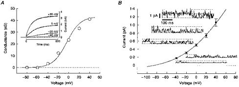
1. Two early signs of human myoblast commitment to fusion are membrane potential hyperpolarization and concomitant expression of a non-inactivating delayed rectifier K+ current, IK(NI). This current closely resembles the outward K+ current elicited by rat ether-à-go-go (r-eag) channels in its range of potential for activation and unitary conductance. 2. It is shown that activation kinetics of IK(NI), like those of r-eag, depend on holding potential and on [Mg2+]o, and that IK(NI), like r-eag, is reversibly inhibited by a rise in [Ca2+]i. 3. Forced expression of an isolated human ether-à-go-go K+ channel (h-eag) cDNA in undifferentiated myoblasts generates single-channel and whole-cell currents with remarkable similarity to IK(NI). 4. h-eag current (Ih-eag) is reversibly inhibited by a rise in [Ca2+]i, and the activation kinetics depend on holding potential and [Mg2+]o. 5. Forced expression of h-eag hyperpolarizes undifferentiated myoblasts from -9 to -50 mV, the threshold for the activation of both Ih-eag and IK(NI). Similarly, the higher the density of IK(NI), the more hyperpolarized the resting potential of fusion-competent myoblasts. 6. It is concluded that h-eag constitutes the channel underlying IK(NI) and that it contributes to the hyperpolarization of fusion-competent myoblasts. To our knowledge, this is the first demonstration of a physiological role for a mammalian eag K+ channel.


Figures

Figure 1
Figure 1. Modulation of IK(NI) by voltage, [Mg2+]o and [Ca2+]i
A, whole-cell current was recorded from a fusion-competent myoblast after 2 days in differentiation medium. Cell was held at −80 mV (steady-state potential), then stepped to potentials ranging between −60 and −140 mV for 200 ms before the final step to the test potential (+40 mV). Recordings were made in a bath medium containing 3 mm Mg2+ and in the presence of 30 μm nifedipine and 2 μm 4-aminopyridine to block other K+ currents. Leak current was estimated in presence of 90 mm TEA and 5 mm Ba2+, and subtracted. Cell capacitance was 12 pF. The relationship between the half-activation time course (I1/2) and the prepulses are illustrated in the inset, in which symbols indicate the means from nine cells and were connected by straight lines. B, recordings from the same cell as in A. Conditions were the same as in A except that Mg2+ was removed from the bath medium; inset, symbols represent the means from seven cells and were connected by straight lines. C, voltage protocol used in A and B. D, simultaneous recordings of whole-cell current during steady state holding at +40 mV (top panel) and [Ca2+]i (lower panel). The fusion-competent myoblast (2 days in differentiation medium) was held at +40 mV for 3 min prior to data acquisition and perfused with extracellular medium containing 3 mm TEA to block large conductance calcium-activated K+ channels (Hamann et al. 1994). Ionomycin (0.5 μm) was added to the extracellular medium and perfused as indicated (horizontal bar). Leak current was estimated by linear extrapolation from the current-voltage relationship from −90 to −60 mV, and subtracted. [Ca2+]i was determined using fura-2. Inset, outward current at +40 mV plotted against [Ca2+]i, during the recovery phase. The continuous line is a Hill equation with IC50 of 144 nm and a Hill coefficient (nH) of 4.5. In five cells, IC50 and nH were 135 ± 28 nm and 3.6 ± 0.3, respectively.
Figure 2
Figure 2. Properties of h-eag current in transfected human undifferentiated myoblasts
A, recordings were performed in cells labelled with GFP 48 h after transfection of undifferentiated myoblasts with the bicistronic h-eag-GFP vector. Whole-cell outward currents were measured at the end of 600 ms steps and leak current subtracted. The leak current was determined in presence of 90 mm TEA and 5 mm Ba2+. Steady-state conductances were plotted against step potentials and were well described by a Boltzmann equation. Cell capacitance was 24 pF. Inset, voltage-dependent activation of h-eag channels at different voltage steps. B, membrane patches (outside-out) were excised from undifferentiated myoblasts transfected with the h-eag-GFP vector. Symbols (▪) represent the mean single-channel amplitudes obtained from all-points amplitude histograms computed from current traces at potentials between −40 and +40 mV (n = 4). Examples of single-channel recordings are shown for each potential. The continuous line is fitted using the Goldman-Hodgkin-Katz equation.
Figure 3
Figure 3. Modulation of h-eagpotassium channels by voltage, [Mg2+]o and [Ca2+]i
A and B, whole-cell outward currents were recorded from an undifferentiated myoblast 48 h after transfection with h-eag-GFP. Data were acquired either in the presence (A) or absence (B) of 3 mm Mg2+. All recordings were performed in the same conditions as for IK(NI), with 30 μm nifedipine and 2 μm 4-aminopyridine added to the bath medium. Leak current was estimated in presence of 90 mm TEA and 5 mm Ba2+, and subtracted. Cell capacitance was 17 pF. Insets, half-activation time course (I1/2) of Ih-eag were plotted against the prepulses. Symbols are the means from four cells and were connected by straight lines. C, voltage protocol used in A and B. D, effect of a rise of [Ca2+]i on Ih-eag recorded from a h-eag-transfected undifferentiated myoblast. The cell was steadily held at −80 mV and then stepped to +40 mV for 400 ms. Ionomycin (0.5 μm) was added to the bath medium and perfused as indicated (horizontal bar). Current amplitudes were measured at the end of 400 ms steps and not corrected for leak current. Cell capacitance was 50 pF. Inset, current traces recorded before (▪), during (♦), and after (*) ionomycin application.
Figure 4
Figure 4. IK(NI), Ih-eag and membrane resting potential of myoblasts
A, whole-cell IK(NI) and resting membrane potentials were measured in fusion-competent myoblasts after 2–3 days in differentiation medium. IK(NI) was measured at +40 mV. Resting membrane potentials were determined in current-clamp mode. Symbols represent the means of current density and resting potential of the indicated number of cells. The dotted line was drawn by eye. B, ▵ represents the means of Ih-eag density and resting potential measured from h-eag-transfected undifferentiated myoblasts, 48 h after transfection. Ih-eag was measured at +40 mV. Parallel measurements were performed on undifferentiated myoblasts that were mock-transfected and ▴ (error bars are not apparent because they are smaller than symbol) represents the means of the resting membrane potential and IK(NI) density.

References

    1. Arcangeli A, Bianchi L, Becchetti A, Faravelli L, Coronnello M, Mini E, Olivotto M, Wanke E. A novel inward-rectifying K+ current with a cell-cycle dependence governs the resting potential of mammalian neuroblastoma cells. The Journal of Physiology. 1995;489:455–471. - PMC - PubMed
    1. Baroffio A, Aubry JP, Kaelin A, Krause RM, Hamann M, Bader CR. Purification of human muscle satellite cells by flow cytometry. Muscle and Nerve. 1993;16:498–505. - PubMed
    1. Bernheim L, Liu J-H, Hamann M, Haenggeli CA, Fischer-Lougheed J, Bader CR. Contribution of a non-inactivating potassium current to the resting potential of fusion-competent human myoblasts. The Journal of Physiology. 1996;493:129–141. - PMC - PubMed
    1. Binggeli R, Weinstein RC. Membrane potentials and sodium channels: hypotheses for growth regulation and cancer formation based on changes in sodium channels and gap junctions. Journal of Theoretical Biology. 1986;123:377–401. - PubMed
    1. Brüggemann A, Stühmer W, Pardo LA. Mitosis-promoting factor-mediated suppression of a cloned delayed rectifier potassium channel expressed in Xenopus oocytes. Proceedings of the National Academy of Sciences of the USA. 1997;94:537–542. - PMC - PubMed

Publication types

MeSH terms

LinkOut - more resources