. 1998 Oct 15;512 ( Pt 2)(Pt 2):365-76.
doi: 10.1111/j.1469-7793.1998.365be.x.

Contrasting Ca2+ channel subtypes at cell bodies and synaptic terminals of rat anterioventral cochlear bushy neurones

Affiliations

Contrasting Ca2+ channel subtypes at cell bodies and synaptic terminals of rat anterioventral cochlear bushy neurones

J M Doughty et al. J Physiol. .

Abstract

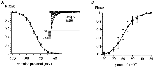
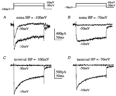
1. Whole-cell patch clamp recordings were made from bushy cells of the anterioventral cochlear nucleus (aVCN) and their synaptic terminals (calyx of Held) in the medial nucleus of the trapezoid body (MNTB). 2. Both high voltage-activated (HVA) and low voltage-activated (LVA) calcium currents were present in acutely dissociated aVCN neurones and in identified bushy neurones from a cochlear nucleus slice. 3. The transient LVA calcium current activated rapidly on depolarization (half-activation, -59 mV) and inactivated during maintained depolarization (half-inactivation, -89 mV). This T-type current was observed in somatic recordings but was absent from presynaptic terminals. 4. On the basis of their pharmacological sensitivity, P/Q-type Ca2+ channels accounted for only 6 % of the somatic HVA, while L-, N- and R-type Ca2+ channels each accounted for around one-third of the somatic calcium current. 5. The divalent permeabilities of these native calcium channels were compared. The Ba2+/Ca2+ conductance ratios of the somatic HVA and LVA channels were 1.4 and 0.7, respectively. The conductance ratio of the presynaptic HVA current was 0.9, significantly lower that that of the somatic HVA current. 6. We conclude that LVA currents are expressed in the bushy cell body, but are not localized to the excitatory synaptic terminal. All of the HVA current subtypes are expressed in bushy cells, but there is a strong polarity to their localization; P-type contribute little to somatic currents but predominate at the synaptic terminal; L-, N- and R-types dominate at the soma, but contribute negligibly to calcium currents in the terminal.


Figures

Figure 1
Figure 1. Calcium currents in somata of dissociated aVCN neurones
A, series of calcium currents evoked in the same neurone from holding potentials of either −80 mV (upper traces) showing HVA currents alone or −120 mV (lower traces) showing both LVA and HVA currents. B, current-voltage relationships for data in A measured at the peak of the inward current; ○, from −120 mV; •, from −80 mV. Charge carrier, 10 mm [Ba2+]o.
Figure 2
Figure 2. Properties of the LVA calcium current
A, inactivation of the LVA current was measured on stepping to a test voltage of −50 mV following 500 ms prepulses to between −120 and −60 mV. Averaged data from 8 cells were fitted with a Boltzmann relationship of the form I/Imax= 1/(1 + exp(V1/2V)/k), giving half-inactivation (V1/2) of −89 mV and a slope (k) of −7 mV. Inset shows example traces from 1 cell. B, activation of the LVA current was examined in 3 neurones. Average data from 3 cells are plotted and fitted to a Boltzmann relationship with half-activation of −60 mV and a slope of 6 mV.
Figure 3
Figure 3. Pharmacology of somatic HVA currents in dissociated aVCN cell bodies
A, pharmacology of the somatic HVA current evoked from a holding potential of −80 mV. The calcium current amplitude is plotted against time with bars showing cumulative application of nifedipine (10 μm), ω-conotoxin GVIA (CTx GVIA; 2 μm) and ω-agatoxin IVA (AgaTx; 200 nm). Example traces are shown on the right. Data were collected using 2 mm [Ca2+]o. B, when applied alone, the block by ω-conotoxin GVIA (1 μm) was partially reversible and voltage independent. For this experiment calcium currents were elicited by ramp depolarizations to +50 mV from a holding potential of −80 mV and using 10 mm [Ba2+]o as the charge carrier. The peak current is plotted against time on the left and example ramps are shown superimposed on the right.
Figure 4
Figure 4. Summary of somatic HVA subtypes
Relative contribution of each current type to the total somatic HVA current (means ± s.e.m.). Data from toxin block experiments using Ba2+ and Ca2+ were pooled (n = 12 for L-, N- and P/Q- type and n = 7 for R-type current).
Figure 5
Figure 5. Comparison of LVA and HVA currents in bushy neurone somata and terminals
Examples of somatic calcium currents (using 2.5 mm [Ca2+]o) are shown in A and B, and synaptic terminal calcium currents (using 1 mm [Ca2+]o) are shown in C and D. A, soma, holding potential (HP) of −100 mV. A transient LVA current was observed when the cell soma was stepped to −50 mV, while a step to −10 mV also elicited a HVA current. B, soma, holding potential of −70 mV (in the same cell as A). The LVA current was inactivated so no current was observed on stepping to −50 mV and only the HVA current was elicited on stepping to −10 mV. C, terminal, holding potential of −100 mV. On stepping to −50 mV no LVA current was evoked, but a HVA current was evoked on stepping to −10 mV. D, terminal, holding potential of −70 mV (in the same terminal as C). The currents were similar to those evoked on stepping from −100 mV.
Figure 6
Figure 6. Ba2+/Ca2+ conductance ratios of somatic and terminal calcium currents
Comparison of the Ba2+/Ca2+ conductance ratio between calcium currents in bushy cell nerve terminals and somata. I–V curves are shown on the left for 1 mm [Ca2+]o (○) and 1 mm [Ba2+]o (•) and example traces are shown on the right. Data are shown for individual cells in each panel; see text for mean values. The I–V data have been fitted with a modified Boltzmann function (see Methods). A, synaptic terminal HVA current. I–V relationship for the terminal P-type calcium current measured from a holding potential of −60 mV. Substitution of Ba2+ for Ca2+ as the charge carrier shifted the half-activation by −4 mV in this terminal with little effect on the peak conductance. B, somatic HVA currents. I–V relationship for the somatic HVA currents evoked from a holding potential of −60 mV. Substitution of Ba2+ for Ca2+ shifted the half-activation by −6 mV in this cell and dramatically potentiated the peak conductance. C, somatic LVA currents. I–V relationships for the same cell as in B, evoked after a 500 ms, −120 mV prepulse to relieve inactivation of the LVA current. Data were fitted with the sum of two Boltzmann equations to account for activation of both the LVA and HVA currents.
Figure 7
Figure 7. ACPD depressed the somatic HVA but not the LVA current
A, combined LVA and HVA currents induced from a holding potential of −120 mV in control (left) and on application of 200 μm 1S,3R-ACPD (right) showing depression of the sustained HVA current. B, the current-voltage relationships for the same data plotted at the peak (circles) and end of the voltage step (squares). Open symbols, control; filled symbols, 200 μm 1S,3R-ACPD. The LVA current amplitude was unaffected, but the HVA current was reduced by ACPD in a voltage-independent manner.

References

    1. Barnes-Davies M, Forsythe ID. Pre- and postsynaptic glutamate receptors at a giant excitatory synapse in rat auditory brainstem slices. The Journal of Physiology. 1995;488:387–406. - PMC - PubMed
    1. Berrow NS, Brice NL, Tedder I, Page KM, Dolphin AC. Properties of cloned rat α1A calcium channels transiently expressed in the COS-7 cell line. European Journal of Neuroscience. 1997;9:739–748. - PubMed
    1. Borst JGG, Helmchen F, Sakmann B. Pre- and postsynaptic whole-cell recordings in the medial nucleus of the trapezoid body of the rat. The Journal of Physiology. 1995;489:825–840. - PMC - PubMed
    1. Borst JGG, Sakmann B. Calcium current during a single action potential in a large presynaptic terminal of the rat brainstem. The Journal of Physiology. 1998;506:143–157. - PMC - PubMed
    1. Bourinet E, Zamponi GW, Stea A, Soong TW, Lewis BA, Jones LP, Yue DT, Snutch TP. The α1E calcium channel exhibits permeation properties similar to low-voltage-activated calcium channels. Journal of Neuroscience. 1996;16:4983–4993. - PMC - PubMed

Publication types

LinkOut - more resources