. 1998 Oct 15;512 ( Pt 2)(Pt 2):435-48.
doi: 10.1111/j.1469-7793.1998.435be.x.

Involvement of PKC-alpha in regulatory volume decrease responses and activation of volume-sensitive chloride channels in human cervical cancer HT-3 cells

Affiliations

Involvement of PKC-alpha in regulatory volume decrease responses and activation of volume-sensitive chloride channels in human cervical cancer HT-3 cells

C Y Chou et al. J Physiol. .

Abstract

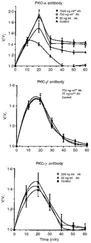
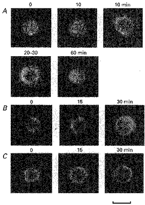
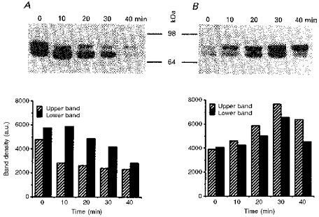
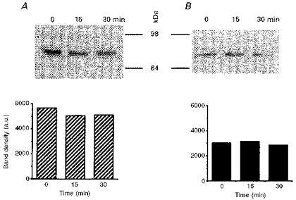
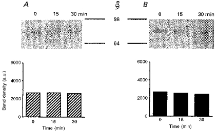
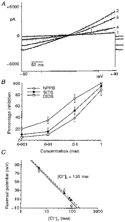
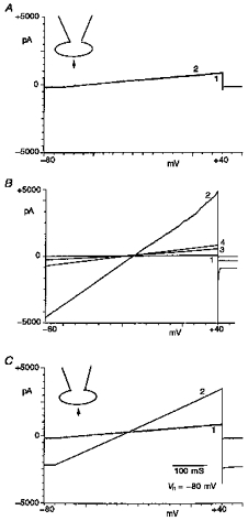
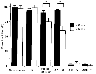
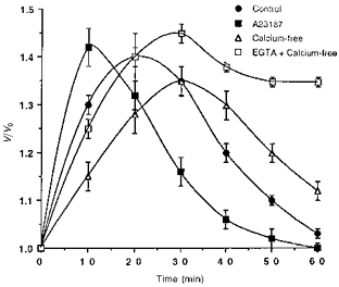
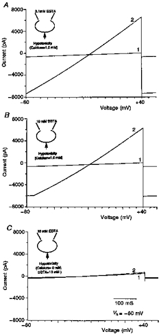
1. The present study was carried out to identify the specific protein kinase C (PKC) isoform involved in regulatory volume decrease (RVD) responses, and to investigate the signal transduction pathways underlying the activation of volume-sensitive chloride channels in human cervical cancer HT-3 cells. The role of Ca2+ in RVD and in the activation of chloride currents was also studied. 2. The time course of RVDs was prolonged by microinjection of PKC-alpha antibody but not by PKC-beta or PKC-gamma antibody, and also by exposure to Ca2+-free medium, in particular when combined with microinjection of EDTA. Immunofluorescence staining showed that hypotonic superfusion evoked the translocation of PKC-alpha to the cell membrane, whereas PKC-beta or PKC-gamma remained unaffected. The translocation of PKC-alpha was observed a few minutes after hypotonic stress, reaching peak intensity at 30 min, and returned to the cytoplasm 60 min after hypotonic exposure. Western blot analyses showed an increased PKC-alpha level in terms of intensity and phosphorylation in the cell membrane, while neither PKC-beta nor PKC-gamma was activated upon hyposmotic challenge. 3. Whole-cell patch-clamp studies demonstrated that neomycin and PKC blockers such as staurosporine and H7 inhibited volume-sensitive chloride currents. The inhibitory effect of neomycin on chloride currents can be reversed by the PKC activator phorbol 12-myristate, 13-acetate (PMA). Moreover, the PKC inhibitor and PKC-alpha antibody, but not PKC-beta or PKC-gamma antibody, significantly attenuated the chloride currents. The activation of volume-sensitive chloride currents were insensitive to the changes of intracellular Ca2+ but required the presence of extracellular Ca2+. 4. Our results suggest the involvement of PKC-alpha and extracellular Ca2+ in RVD responses and the activation of volume-sensitive chloride channels in HT-3 cells.


Figures

Figure 1
Figure 1. Effects of isoform-specific PKC antibodies on the time course of RVD in HT-3 cells
Examples of time course of volume changes in HT-3 cells following superfusion with 210 mosmol l−1 hypotonic bath solution are shown. The isoform-specific PKC antibody was delivered by microinjection. Ten minutes after microinjection, hypotonic solution was perfused. The y-axis (V/V0) depicts the cell volume at the indicated times divided by the cell volume at zero time. Each point represents mean ± s.e.m. (n = 15 cells) from three separate experiments.
Figure 2
Figure 2. Immunofluorescence staining of PKC isoforms in HT-3 cells
These representative pictures show the intracellular localization of PKC-α (A),-β (B) and -γ (C), respectively, in isotonic conditions at various time periods after 210 mosmol l−1 hypotonic solution perfusion. The observations showing the effects of hyposmolarity on β- and γ-PKC are shown 15 and 30 min after hypotonic solution perfusion. Scale bar, 10 μm. For details, see Methods.
Figure 3
Figure 3. Western blot and quantification of PKC-α in HT-3 cells
HT-3 cells were exposed to isotonic solution (290 mosmol l−1) and then lysed immediately or after hypotonic solution (210 mosmol l−1) perfusion for 10–40 min. After separation of cytosolic (A) and particulate (B) fractions, 15 μg of each protein from the two fractions was denatured in SDS lysis buffer and loaded into 10 % SDS-polyacrylamide gels. Blotting and immunodetection with PKC-α antibody were then carried out. Histograms represent the densitometric reading of the corresponding bands. For details of the assay see Methods. A typical experiment was shown here, which had been repeated five times with comparable results. The data in lane 1, panel B has been cut and then pasted from a different lane of the same film. a.u., arbitrary units.
Figure 4
Figure 4. Western blot and quantification of PKC-β in HT-3 cells
HT-3 cells were exposed to isotonic solution (290 mosmol l−1) and then lysed immediately or after hypotonic solution (210 mosmol l−1) perfusion for 15 or 30 min. After separation of cytosolic (A) and particulate (B) fractions, 15 μg of each protein from the two fractions was denatured in SDS lysis buffer and loaded into 10 % SDS-polyacrylamide gels. Blotting and immunodetection with PKC-β antibody were then carried out. Histograms represent the densitometric reading of the corresponding bands. For details of the assay see Methods. A typical experiment is shown here, which had been repeated five times with comparable results.
Figure 5
Figure 5. Western blot and quantification of PKC-γ in HT-3 cells
HT-3 cells were exposed to isotonic solution (290 mosmol l−1) and then lysed immediately or after hypotonic solution (210 mosmol l−1) perfusion for 15 or 30 min. After separation of cytosolic (A) and particulate (B) fractions, 15 μg of each protein from the two fractions was denatured in SDS lysis buffer and loaded into 10 % SDS-polyacrylamide gels. Blotting and immunodetection with PKC-γ antibody were then carried out. Histograms represent the densitometric reading of the corresponding bands. For details of the assay see Methods. A typical experiment is shown here, which had been repeated five times with comparable results.
Figure 6
Figure 6. Characterization of volume-sensitive chloride currents in HT-3 cells
A, a Cl channel blocker, NPPB, with a dose-dependent manner inhibited the volume-sensitive Cl current in HT-3 cells. The membrane potential was held at −80 mV and the ramp command pulse from −80 to +40 mV was employed. 1, basal membrane current recorded under isotonic conditions (290 mosmol l−1); 2, current recorded after perfusion with hypotonic solution (210 mosmol l−1); 3 and 4, currents recorded after adding 10 or 100 μm NPPB to the perfusing hypotonic solution. B, dose-response relationships of Cl channel blockers for the percentage of inhibition of volume-sensitive Cl current measured at +40 mV. Each point is mean ± s.e.m. (n = 6). C, the reversal potential for the volume-sensitive Cl current at various extracellular Cl concentrations ([Cl]o). The line was well fitted by the linear regression analysis. The dashed line represents the theoretical values of reversal potential in different Cl concentrations estimated by the Goldman-Hodgkin-Katz equation.
Figure 7
Figure 7. Effects of neomycin, staurosporine and the combined use of neomycin and PMA on volume-sensitive chloride currents in HT-3 cells
A, effect of neomycin, a blocker of phospholipase C, on volume-sensitive Cl current in HT-3 cells. 1, basal membrane current recorded in isotonic conditions; 2, current recorded after perfusion with hypotonic solution when 1 mm neomycin was added to the pipette solution. B, effect of staurosporine, a blocker of protein kinase C, on volume-sensitive Cl current in HT-3 cells. 1, basal membrane current recorded in isotonic conditions; 2, current recorded after perfusion with hypotonic solution; 3, current recorded after perfusing solution was shifted from hypotonicity to isotonicity; 4, current recorded after adding 1 μm staurosporine to the perfusing hypotonic solution. C, interaction of neomycin and PMA on volume-sensitive Cl current in HT-3 cells. 1, current recorded after perfusing with hypotonic solution when 1 mm neomycin was added to the pipette solution; 2, current recorded after adding 0.1 μm PMA to the hypotonic solution and 1 mm neomycin to the pipette solution; Vh, holding potential.
Figure 8
Figure 8. Effects of PKC blockers and isoform-specific PKC antibodies on volume-sensitive chloride currents in HT-3 cells
The effects of PKC blockers such as staurosporine and H7, PKC peptide inhibitors, and isoform-specific PKC antibodies on volume-sensitive chloride currents measured at −80 and +40 mV. The y-axis is the percentage inhibition of volume-sensitive chloride currents induced by each blocker. Each bar represents the mean ± s.e.m. (n = 16); * significance level of P < 0.05 (paired t test). Peptide inhibitor is the PKC inhibitor as mentioned in Methods.
Figure 9
Figure 9. Role of calcium on cell volume regulation of HT-3 cells on exposure to 210 mosmol l−1 hypotonic stress
The y-axis (V/V0) depicts the cell volume at the indicated times divided by the cell volume at zero time. Each point represents mean ± s.e.m. (n = 20 cells) from three separate experiments. A23187 (10 μm) was added to the hypotonic medium. Calcium-free medium was the hypotonic medium without addition of calcium. EGTA was transferred into cells by microinjection. EGTA at 1 mm had minimal effect on RVD process (data not shown).
Figure 10
Figure 10. Effect of calcium depletion on the activation of volume-sensitive Cl current in HT-3 cells
A, 1, basal membrane current recorded in isotonic conditions. 2, current recorded in the hypotonic perfusing solution with 0.1 mm EGTA in the pipette solution. B, 1, basal membrane current recorded in isotonic conditions; 2, current recorded in the hypotonic perfusing solution with 10 mm EGTA in the pipette solution. C, 1, basal membrane current recorded in isotonic conditions; 2, current recorded in the calcium-free hypotonic solution with 10 mm EGTA in the pipette solution.

References

    1. Borner C, Filipuzzi I, Wartmann M, Eppenberger U, Fabbro D. Biosynthesis and post-translational modifications of protein kinase C in human breast cancer cells. Journal of Biological Chemistry. 1989;264:13902–13909. - PubMed
    1. Chang WC, Ning CC, Lin MT, Huang JD. Epidermal growth factor enhances a microsomal 12-lipoxygenase activity in A431 cells. Journal of Biological Chemistry. 1992;267:3657–3666. - PubMed
    1. Chapline C, Mousseau B, Ramsay K, Duddy S, Li Y, Kiley SC, Jaken S. Identification of a major kinase C-binding protein and substrate in rat embryo fibroblasts: decreased expression in transformed cells. Journal of Biological Chemistry. 1996;271:6417–6422. . - DOI - PubMed
    1. Chou CY, Shen MR, Chen TM, Huang KE. Volume-activated taurine transport is differentially activated in human cervical cancer HT-3 cells, but not activated in HPV-immortalized Z 183A and normal cervical epithelial cells. Clinical and Experimental Pharmacology and Physiology. 1997;24:935–939. - PubMed
    1. Chou CY, Shen MR, Wu SN. Volume-sensitive chloride channels associated with human cervical carcinogenesis. Cancer Research. 1995;55:6077–6083. - PubMed

Publication types

LinkOut - more resources