. 1998 Oct 1;12(19):3008-19.
doi: 10.1101/gad.12.19.3008.

Premature senescence involving p53 and p16 is activated in response to constitutive MEK/MAPK mitogenic signaling

Affiliations

Premature senescence involving p53 and p16 is activated in response to constitutive MEK/MAPK mitogenic signaling

A W Lin et al. Genes Dev. .

Abstract

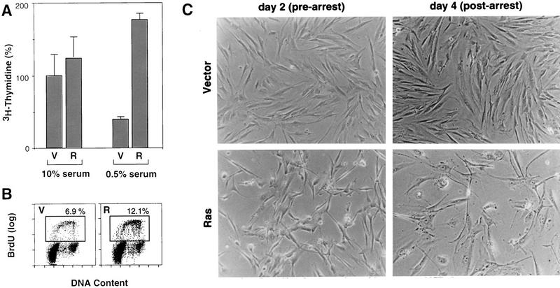
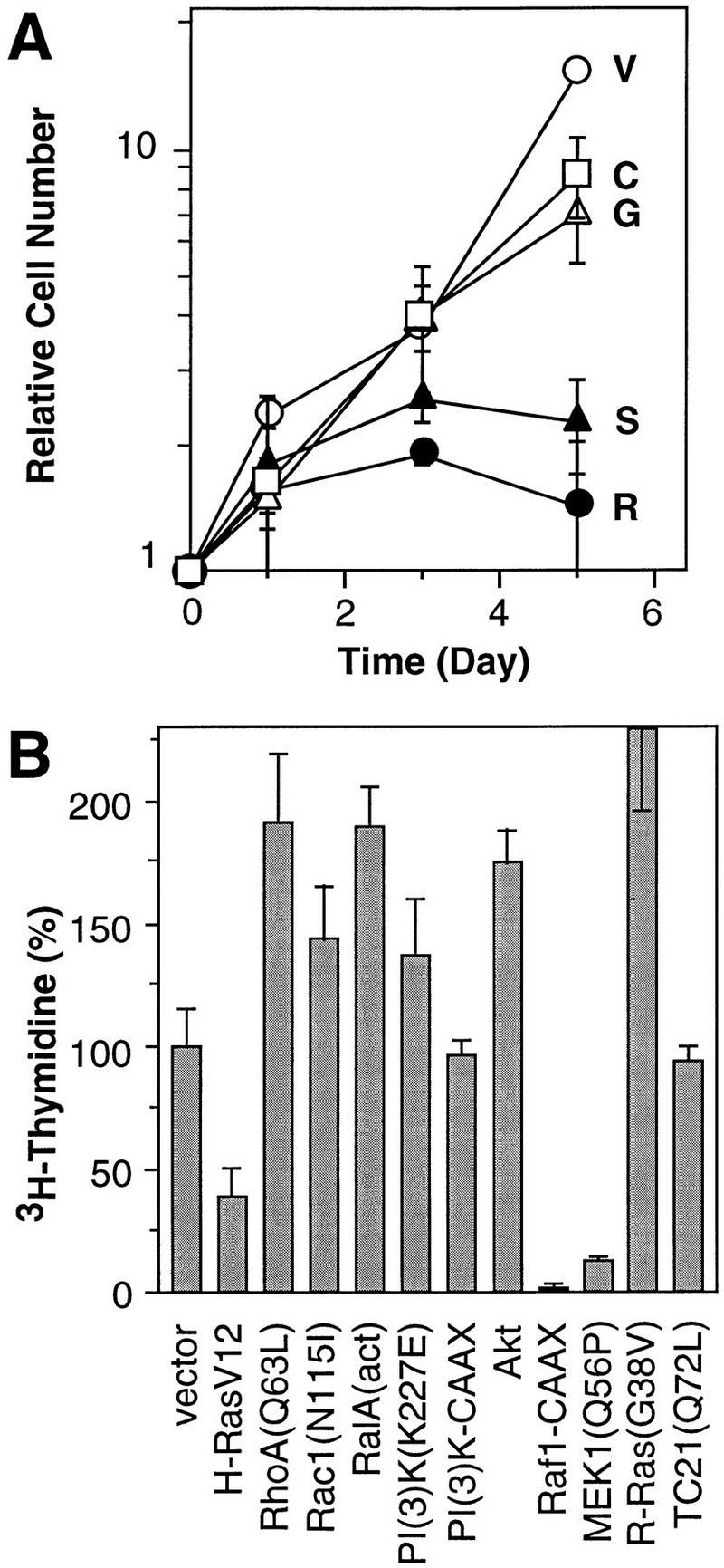
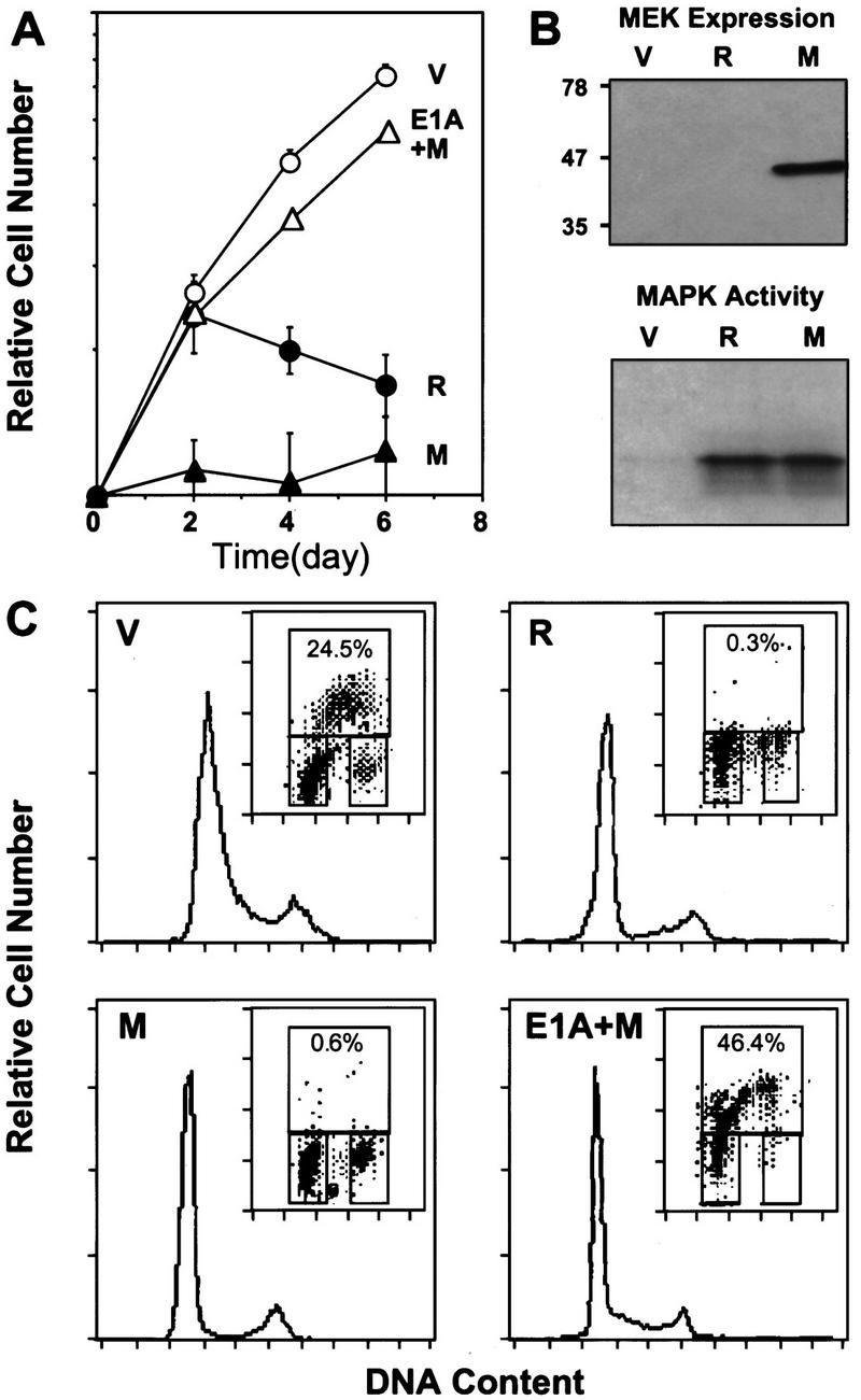
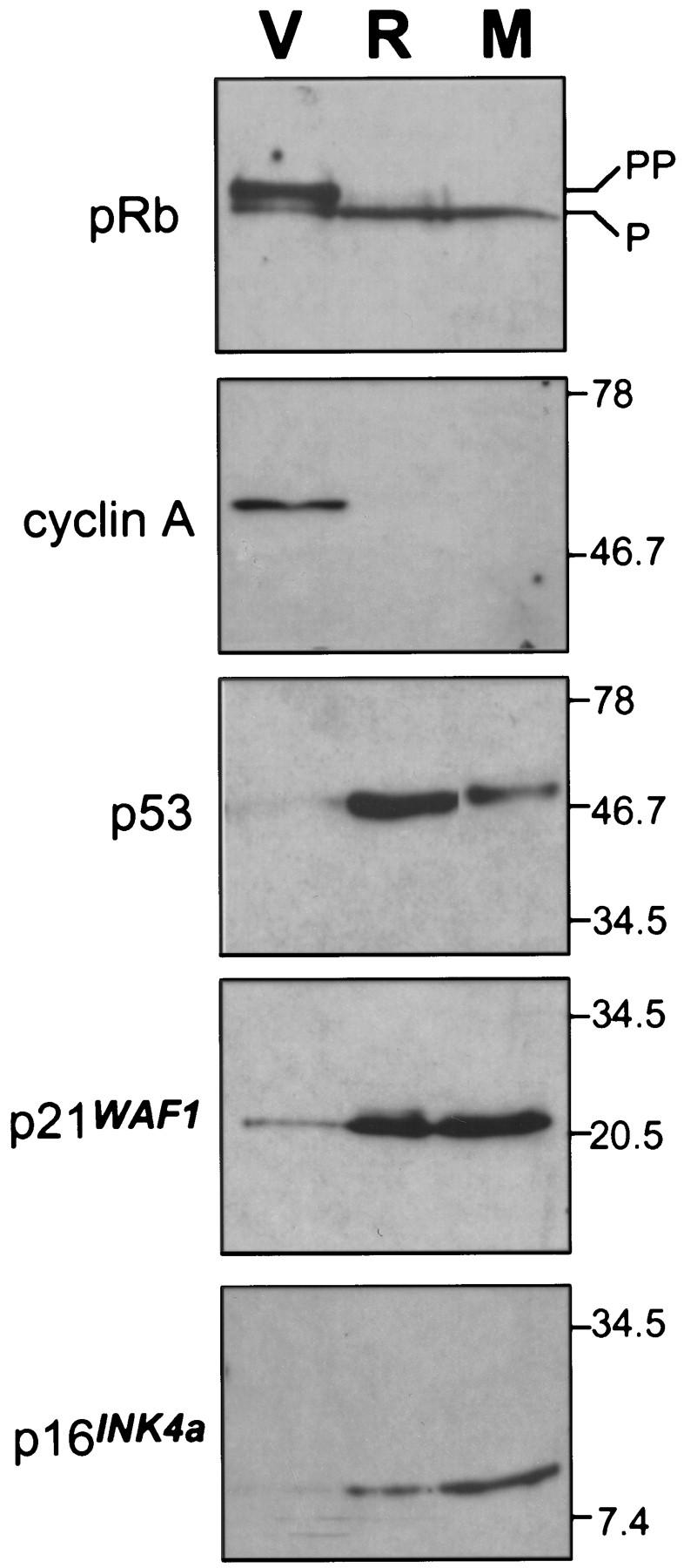
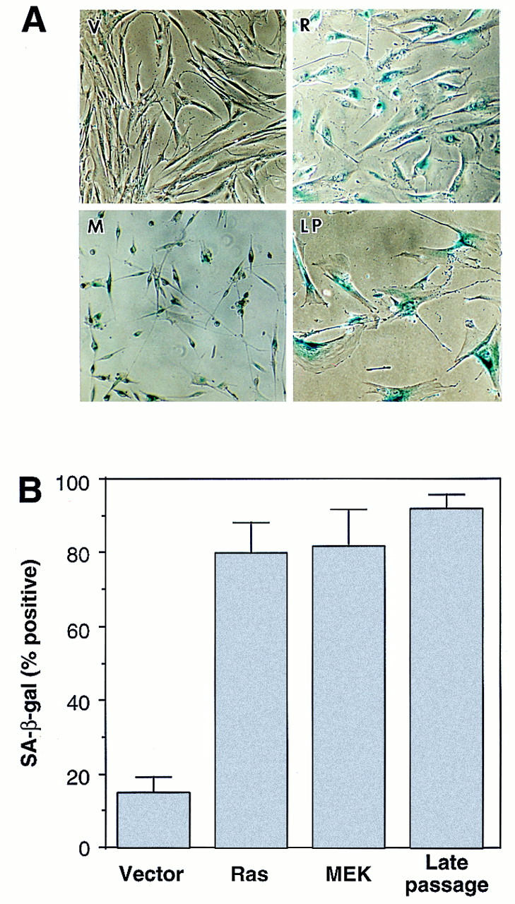
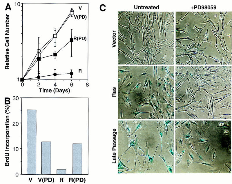
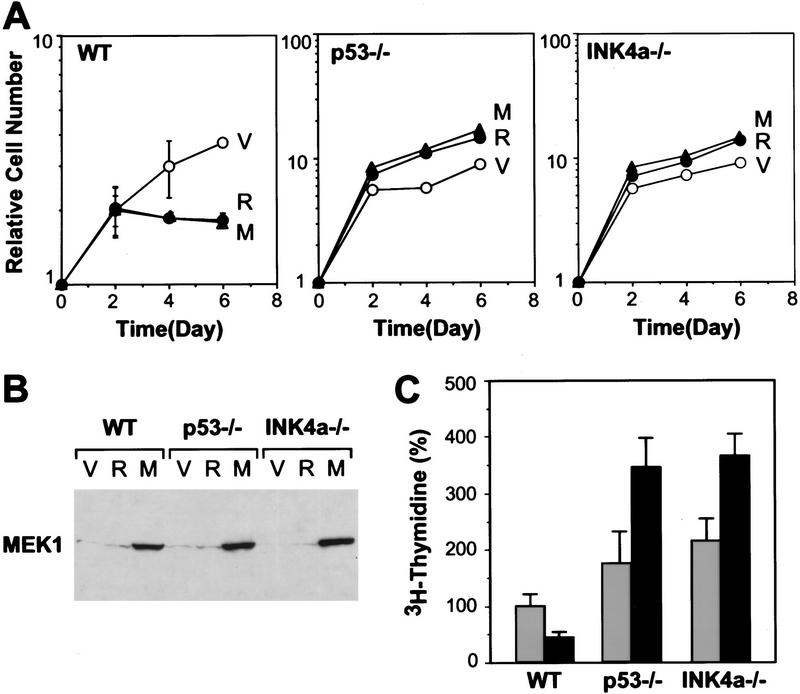
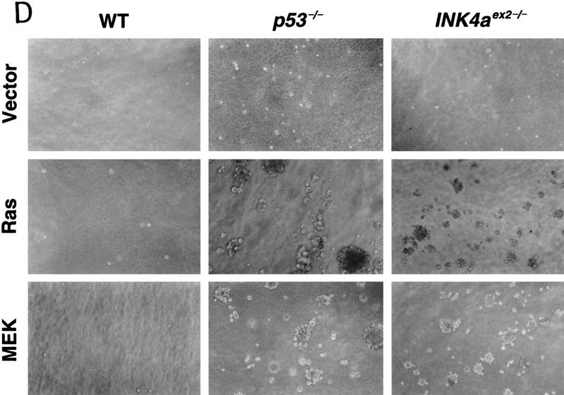
Oncogenic Ras transforms immortal rodent cells to a tumorigenic state, in part, by constitutively transmitting mitogenic signals through the mitogen-activated protein kinase (MAPK) cascade. In primary cells, Ras is initially mitogenic but eventually induces premature senescence involving the p53 and p16(INK4a) tumor suppressors. Constitutive activation of MEK (a component of the MAPK cascade) induces both p53 and p16, and is required for Ras-induced senescence of normal human fibroblasts. Furthermore, activated MEK permanently arrests primary murine fibroblasts but forces uncontrolled mitogenesis and transformation in cells lacking either p53 or INK4a. The precisely opposite response of normal and immortalized cells to constitutive activation of the MAPK cascade implies that premature senescence acts as a fail-safe mechanism to limit the transforming potential of excessive Ras mitogenic signaling. Consequently, constitutive MAPK signaling activates p53 and p16 as tumor suppressors.


Figures

Figure 1
Figure 1
Oncogenic Ras is initially mitogenic in primary fibroblasts. (A) [3H]thymidine incorporation assay using IMR90 cell populations containing empty vector (V) or H–RasV12 (R). Twenty-four hours postinfection, 2 × 14 of the indicated cells were plated and grown in medium containing 10% FBS or 0.5% FBS for 24 hr followed by a 3H]thymidine pulse. Values were normalized to those obtained from control populations in 10% serum; the average and standard deviation of three measures are shown. (B) Cell-cycle analysis of IMR90 cell populations containing empty vector (V) or H–RasV12 (R) in low-serum conditions. Twenty-four hours postinfection, cells were transferred to 0.5% FBS-containing medium, 24 hr later, the cells werepulsed with BrdU for 4 hr; and analyzed for BrdU incorporation and DNA content by two-color flow cytometry (see Materials and Methods). The box represents cells incorporating BrdU (S-phase); the percentage of BrdU positive cells are indicated. (C) Representative photomicrographs of IMR90 cells transduced with empty vector or a H–RasV12-expressing construct. Cells were transduced with retroviruses containing either empty vector or H-RasV12; the photomicrographs were taken at day 2 postselection (prearrest) and day 4 postselection (postarrest) according to the time scheme described in Materials and Methods.
Figure 2
Figure 2
Effect of Ras-dependent signaling pathways on proliferation. (A) Representative growth curves corresponding to the indicated IMR90 cell populations containing empty vector (V, ○), H-RasV12 (R, •) H-RasV12/S35 (S, ▴), H-RasV12/G37 (G, ▵), H-RasV12/C40 (C, □). Each value was determined in triplicate and normalized to the cell number at day 0. (B) Representative [3H]thymidine incorporation assay of IMR90 cell populations expressing various Ras-related proteins, Ras effectors or Ras downstream components (see Materials and Methods). The values were normalized to those obtained from cells containing a control vector; the average and standard deviation of three measures are shown.
Figure 3
Figure 3
Activated MEK induces cell-cycle arrest in human diploid fibroblasts. (A) Representative growth curves corresponding to IMR90 cell populations transduced with empty vector (V, ○), H–RasV12 (R, •), MEK1Q56P (M, ▴) or E1A+MEK1Q56P (E1A+M, ▵). Each value was determined in triplicate and normalized to the cell number at day 0. (B) Expression of ectopic MEK1Q56P in retrovirally infected IMR90 cells was verified by immunoblot analysis at day 4 postselection. MAP kinase activity in cell populations at day 2 postselection was measured by immunoprecipitation of ERK2, followed by a kinase assay using [γ-32P]ATP and myelin basic protein as substrate (see Materials and Methods). (C) Cell-cycle analysis of the indicated cell populations (day 6 postselection) as determined by BrdU incorporation and DNA-content analysis (see Materials and Methods). The histogram displays the DNA profile of the indicated cell population as measured by propidium iodide. (Insets) The upper box indicates cells incorporating BrdU (S phase); the lower-left box displays G0/G1 cell population; the lower-right box indicates cells in G2/M.
Figure 4
Figure 4
Activated MEK induces accumulation of p53 and p16. Immunoblots of cell-cycle regulatory proteins in lysates from cells containing empty vector (V), H–RasV12 (R), or MEKQ56P (M) at day 4 postselection.
Figure 5
Figure 5
MEK activates premature senescence. (A) Photomicrographs of IMR90 cell populations containing an empty vector (V), H–RasV12 (R), or MEK1Q56P (M) stained for SA-β-galactosidase activity (pH 6.0) at day 6 postselection. Late-passage IMR90 cells (LP) having undergone replicative senescence are shown for comparison. (B) Percentage of cells positive for SA-β-galactosidase 6 days after drug selection. Cells (∼ 200) from each cell population were scored; the average and standard deviation of data from at least three separate experiments are shown.
Figure 6
Figure 6
MEK is required for Ras-induced cell-cycle arrest and premature senescence. (A) Representative growth curves documenting the effects of PD98059 on Ras-induced growth arrest. IMR90 cells were infected with an empty vector- or an H-RasV12-expressing retrovirus. Immediately after selection, cell populations were treated daily with medium containing 50 μm PD98059 or 0.25% DMSO (carrier). Each value was determined in triplicate and normalized to the cell number at day 0. (B) Cell populations containing an empty vector (V) or expressing H-RasV12 (R) were treated daily with PD98059 (PD). On day 6 postselection, cells were pulsed with BrdU and analyzed for BrdU incorporation by flow cytometry. The calculated percentage of BrdU-positive cells is shown. (C) Photomicrographs of control and PD98059-treated cell populations stained for SA-β-gal activity on day 6 postselection. The percentage of positive cells in each population was 9% (vector); 12.5% (vector + PD98059); 80% (Ras); 15.4% (Ras + PD98059); 90% (late passage); 91% (late passage + PD98059).
Figure 7
Figure 7
Inactivation of either p53 or INK4aex2 reveals the mitogenic and transforming activity of activated MEK. (A) Representative growth curves corresponding to wild-type (WT), p53-null (p53−/−) or p16-null (INK4aex2−/−) MEFs transduced with empty vector (V), H-RasV12 (R), or MEK1Q56P (M) expressing retroviruses. Each value was determined in triplicate and normalized to the cell number at day 0. (B) MEK1 expression in the indicated cell populations was determined by immunoblotting on day 4 postselection. (C) [3H]thymidine incorporation in the indicated cell populations containing empty vector (gray bars) or MEK1Q56P (black bars). On day 6 postselection, cells were transferred to 0.5% FBS-containing medium for 36 hr, followed by a 24-hr pulse with [3H]thymidine. Each value was determined in triplicate and normalized to the value obtained for wild-type cells containing the empty vector. (D) Analysis of anchorage-independent growth of the indicated MEF populations. Immediately following selection for virus-transduced populations, ∼2 × 14 cells were plated in soft agar (see Materials and Methods). Photomicrographs were taken 3 weeks after plating.
Figure 7
Figure 7
Inactivation of either p53 or INK4aex2 reveals the mitogenic and transforming activity of activated MEK. (A) Representative growth curves corresponding to wild-type (WT), p53-null (p53−/−) or p16-null (INK4aex2−/−) MEFs transduced with empty vector (V), H-RasV12 (R), or MEK1Q56P (M) expressing retroviruses. Each value was determined in triplicate and normalized to the cell number at day 0. (B) MEK1 expression in the indicated cell populations was determined by immunoblotting on day 4 postselection. (C) [3H]thymidine incorporation in the indicated cell populations containing empty vector (gray bars) or MEK1Q56P (black bars). On day 6 postselection, cells were transferred to 0.5% FBS-containing medium for 36 hr, followed by a 24-hr pulse with [3H]thymidine. Each value was determined in triplicate and normalized to the value obtained for wild-type cells containing the empty vector. (D) Analysis of anchorage-independent growth of the indicated MEF populations. Immediately following selection for virus-transduced populations, ∼2 × 14 cells were plated in soft agar (see Materials and Methods). Photomicrographs were taken 3 weeks after plating.

References

    1. Alcorta DA, Xiong Y, Phelps D, Hannon G, Beach D, Barrett JC. Involvement of the cyclin-dependent kinase inhibitor p16 (INK4a) in replicative senescence of normal human fibroblasts. Proc Natl Acad Sci. 1996;93:13742–13747. - PMC - PubMed
    1. Alessi DR, Cuenda A, Cohen P, Dudley DT, Saltiel AR. PD 098059 is a specific inhibitor of the activation of mitogen-activated protein kinase kinase in vitro and in vivo. J Biol Chem. 1995;270:27489–27494. - PubMed
    1. Amati B, Brooks MW, Levy N, Littlewood TD, Evan GI, Land H. Oncogenic activity of the c-Myc protein requires dimerization with Max. Cell. 1993;72:233–245. - PubMed
    1. Atadja P, Wong H, Garkavtsev I, Veillette C, Riabowol K. Increased activity of p53 in senescing fibroblasts. Proc Natl Acad Sci. 1995;92:8348–8352. - PMC - PubMed
    1. Bottorff D, Stang S, Agellon S, Stone JC. RAS signalling is abnormal in a c-raf1 MEK1 double mutant. Mol Cell Biol. 1995;15:5113–5122. - PMC - PubMed

Publication types

MeSH terms

Substances

LinkOut - more resources