. 1998 Oct 1;12(19):3084-95.
doi: 10.1101/gad.12.19.3084.

Synergistic cooperation of TFE3 and smad proteins in TGF-beta-induced transcription of the plasminogen activator inhibitor-1 gene

Affiliations

Synergistic cooperation of TFE3 and smad proteins in TGF-beta-induced transcription of the plasminogen activator inhibitor-1 gene

X Hua et al. Genes Dev. .

Abstract

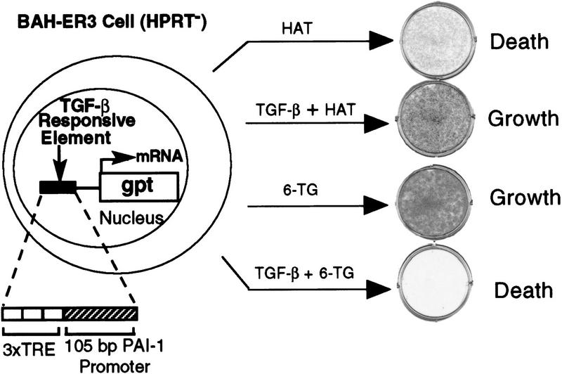
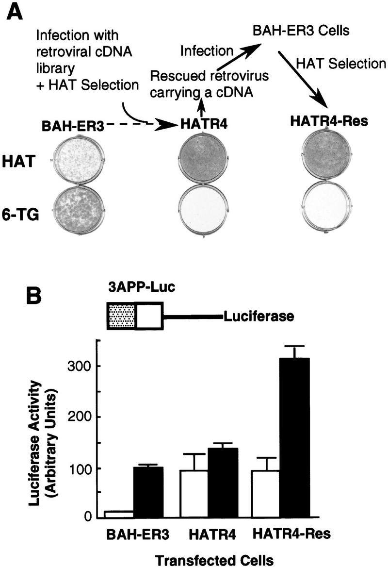
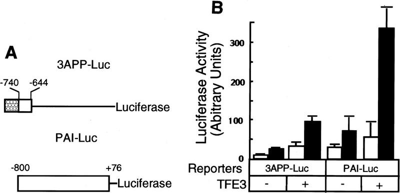
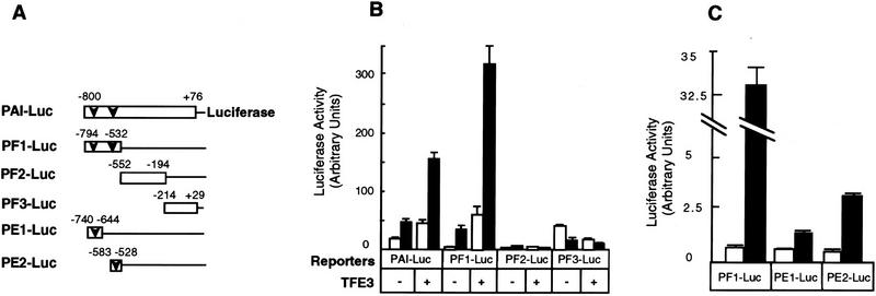
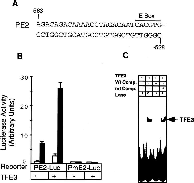
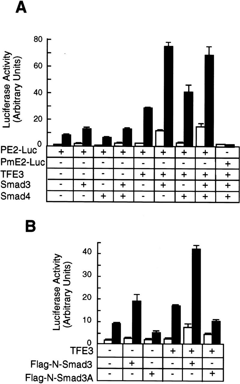
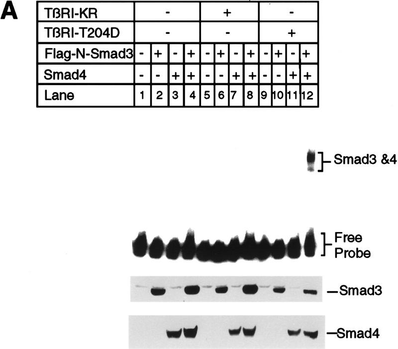
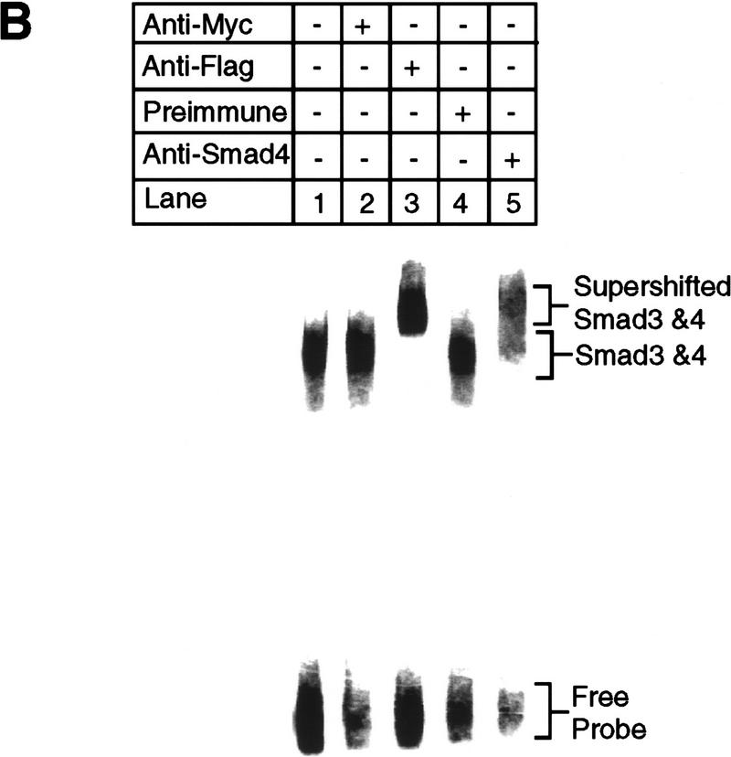
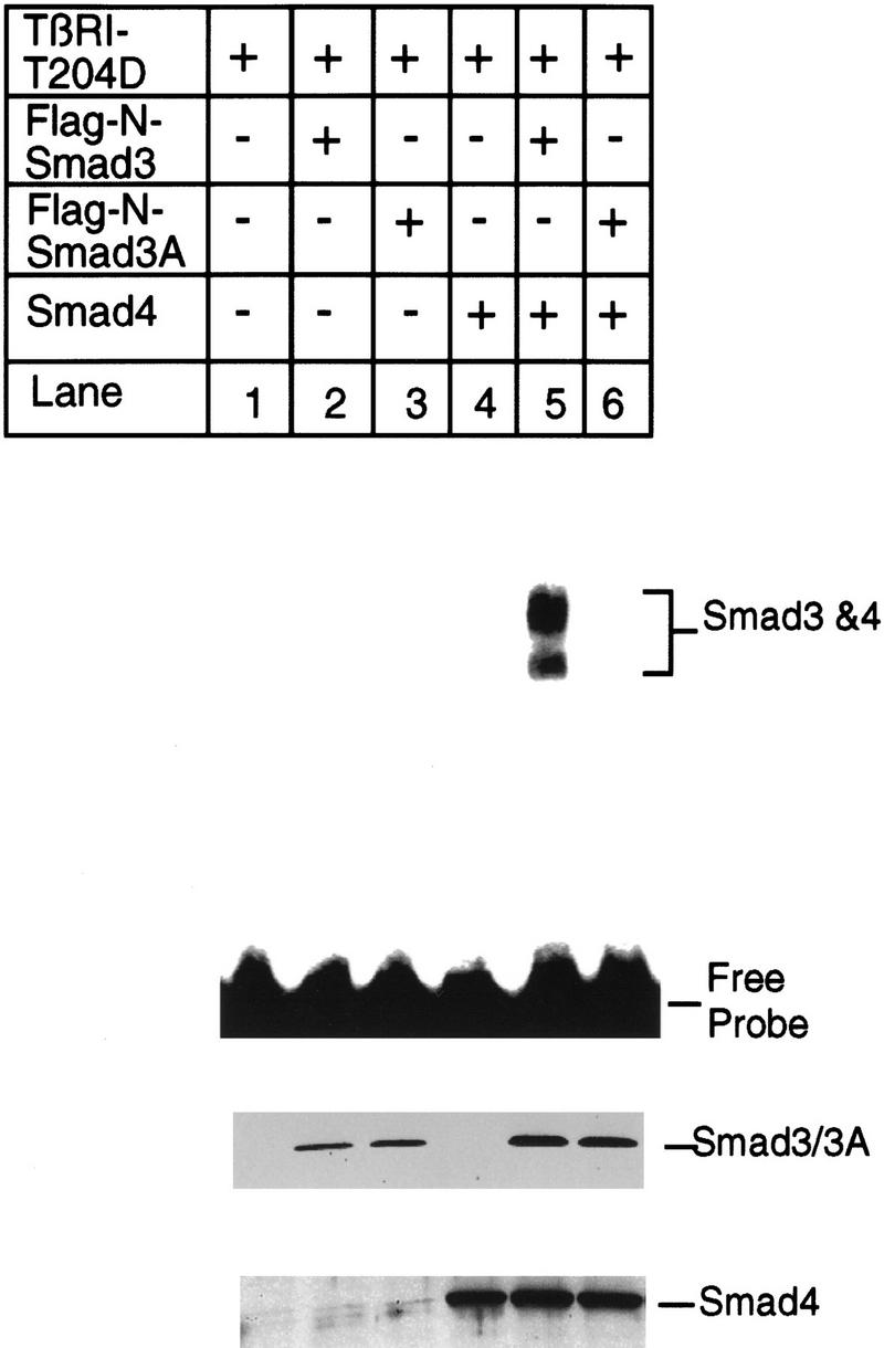
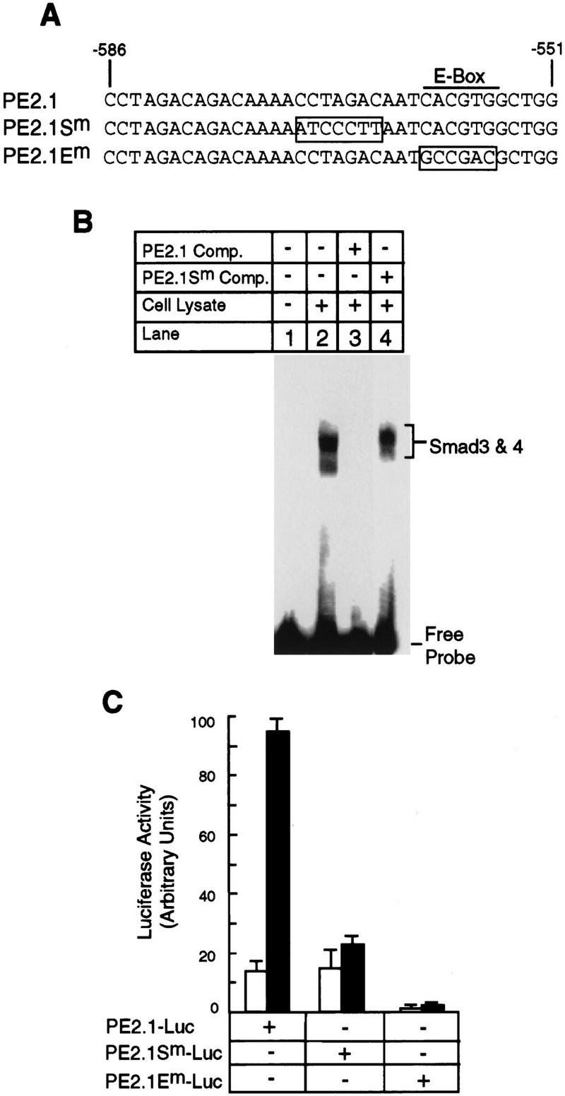
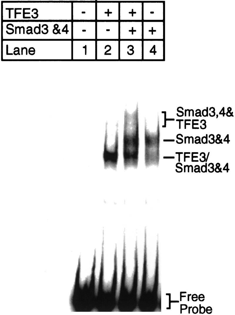
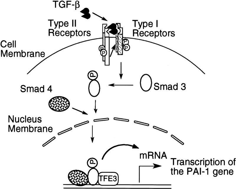
Members of the TGF-beta superfamily influence a broad range of biological activities including stimulation of wound healing and inhibition of cell growth. TGF-beta signals through type I and II receptor serine/ threonine kinases and induces transcription of many genes including plasminogen activator inhibitor-1 (PAI-1). To identify proteins that participate in TGF-beta-induced gene expression, we developed a novel retrovirus-mediated expression cloning strategy; and using this approach, we established that transcription factor microE3 (TFE3) is involved in TGF-beta-induced activation of the PAI-1 promoter. We showed that TFE3 binds to an E-box sequence in PE2, a 56-bp promoter fragment of the PAI-1 promoter, and that mutation of this sequence abolishes both TFE3 binding as well as TGF-beta-dependent activation. TFE3 and Smad3 synergistically activate the PE2 promoter and phosphorylated Smad3 and Smad4 bind to a sequence adjacent to the TFE3-binding site in this promoter. Binding of both TFE3 and the Smad proteins to their cognate sequences is indispensable for TGF-beta-inducible activation of the PE2 promoter. Hence, TFE3 is an important transcription factor in at least one TGF-beta-activated signal transduction pathway.


Figures

Figure 1
Figure 1
TGF-β-regulated growth of BAH-ER3 cells in the presence of drug selection. On day 0, BAH-ER3 cells were seeded at a density of 5 × 104 cells/well in a six-well plate in DMEM containing 10% fetal calf serum, 100 U/ml penicillin and 100 μg/ml streptomycin. On day 1, the cells were switched to medium with 1× HAT or 6-TG (30 μg/ml) with or without 200 pm TGF-β as indicated. On day 9, the growing cells were stained with crystal violet. (TRE) Phorbol ester TPA response element; (gpt) guanosine phosphoribosyl transferase.
Figure 2
Figure 2
Isolation of a HAT-resistant cell clone that activates the TGF-β-inducible 3TP promoter in the absence of TGF-β. (A) After infection of BAH-ER3 cells with a retroviral cDNA library, the cells were grown in HAT medium for 2 weeks. A HAT-resistant clone, HATR4, was isolated, and then infected with wild-type Moloney retrovirus; the supernatant containing the rescued retrovirus was used to infect normal BAH-ER3 cells as described in Materials and Methods. Infected cells were also subjected to HAT selection and the resulting HAT-resistant cells, HATR4–Res cells, were isolated. BAH-ER3, HATR4, and HATR4–Res cells were plated at 5 × 104 cells/well in a 6-well plate; HAT medium or 6-TG medium was added as indicated, and cells were incubated for 10 days in the absence of TGF-β. Growing cells were stained with crystal violet. (B) On day 0, 105 cells were plated in each well of a 12-well plate. On day 2, the cells in each well were transfected with 2 μg of 3APP–Luc DNA and 0.5 μg of pSVβ. After overnight culture the cells were switched to serum-free medium with (█) or without (□) 200 pm TGF-β as indicated, and then incubated for 20 hr before being harvested for luciferase and β-galactosidase assays as described in Materials and Methods. Luciferase activities, plotted in arbitrary units, have been normalized to β-galactosidase activity. Each value represents an average of duplicate samples, and the error bar denotes the standard deviation of the duplicates.
Figure 3
Figure 3
TFE3 activates the PAI-1 promoter in a TGF-β-dependent fashion. (A) A diagram of the luciferase reporter genes driven by various promoters. The three tandem repeats of a 7-bp AP1-binding site [(stippled box) TGA(G/C)TCA] separated by an XbaI site were inserted upstream of −740 to −644 fragment of the PAI-1 promoter (open box) in the 3APP–Luc construct. Hence, the short AP1-binding sequence was used to replace the 32-bp fragment containing an AP1-binding sequence in the 3TP promoter (Wrana et al. 1992) to eliminate the potential influence of sequences other than the AP1-binding site. (B) Activation of PAI–Luc expression by TFE3 is dependent on TGF-β. BAH-ER3 cells were transfected as described in Fig. 2B. Each well received 1.5 μg of reporter gene, 0.5 μg of pSVβ, and also 1.0 μg of plasmid encoding TFE3 as indicated. The total amount of DNA per well was adjusted to 3.0 μg. Transfected cells were treated with (█) or without (□) TGF-β, and processed for both luciferase and β-galactosidase assays as described in Fig. 2B.
Figure 4
Figure 4
Identification of small subfragments, PE1 and PE2, of the PAI-1 promoter that are activated by TFE3 and TGF-β. (A) A diagram of the reporter constructs. (Open bars) PAI-1 promoter; (arrowhead) E-box sequence (CACGTG). BAH-ER3 cells (B) and Hep G2 cells (C) were transfected and the luciferase and β-galactosidase assays were carried out as detailed in Fig. 3B. (□) Without TGF-β; (█) with TGF-β.
Figure 5
Figure 5
The E-box sequence is essential for TFE3-mediated and TGF-β-dependent activation of the PE2 fragment of the PAI-1 promoter. (A) The sequence of the PE2 promoter. (B) Hep G2 cells were transfected with 1 μg of PE2–Luc or PmE2–Luc DNA (CACGTG → acCGac), 1 μg of pSVβ, and also 1.0 μg of plasmid encoding TFE3 as indicated. Transfected cells were treated with (█) or without (□) TGF-β, processed, and assayed as described in Fig. 3B. (C) TFE3 was synthesized in vitro from pET–TFE3 by the TNT T7 Coupled Reticulocyte Lysate System (Promega). Gel-shift reactions contained 3 μl of the in vitro TFE3 translation reaction and 1 μl of (4 × 103 cpm) of 32P-labeled PE2 DNA probe. Reactions in lanes 3 and 4 contained a 50-fold excess of wild type or mutant PE2 oligonucleotides, respectively.
Figure 6
Figure 6
Synergy between TFE3 and Smad3 in the activation of the PE2 promoter. (A) Hep G2 cells were transfected as described in the legend to Fig. 5A. The following plasmids were used in transfection as indicated: 0.5 μg of plasmid encoding TFE3, 1 μg of plasmid encoding Smad3, and 1 μg of plasmid encoding Smad4; every well received 1 μg of PE2–Luc and 0.2 μg of pCMV–β-gal. The total amount of DNA was adjusted to 3.7 μg per well with pcDNA3. (B) Hep G2 cells were transfected with the following plasmids: 0.5 μg of TFE3, 1 μg of Flag-N–Smad3 or Flag-N–Smad3A; every well received 1 μg of PE2–Luc and 0.2 μg of pCMV-β. The total amount of DNA per well was adjusted to 3.7 μg with a control plasmid, pEXL–GFP. The cells were transfected, treated with (█) or without (□) TGF-β, and assayed as described for panel A.
Figure 7
Figure 7
Smad3 and Smad4 together bind to the PE2.1 element of the PAI-1 promoter. (A) Bosc23 cells were transfected as described in Materials and Methods; cells in each dish were transfected with 2 μg of plasmid encoding Flag-N-Smad3 or Smad4 (pEXL–Smad4), and 1 μg of plasmid encoding TβRI-KR (pCMV5–TβRI–KR) or TβRI-T204D (pCMV5-TβRI-T204D) as indicated. The total amount of DNA for each dish was adjusted to 5 μg with pEXL–GFP. The gel-shift assay at top was carried out with the 32P-labeled probe and 1 μl of cell lysate as described in Materials and Methods, and exposed to a Fuji PhosphorImager plate. The minor lower band in lane 12 at top probably represents Smad3 and Smad4 protein binding to only one of the two tandem repeats of the PE2.1 element. (Middle, bottom) Immunoblots with 5 μl (150 μg of proteins) of cell lysates in each lane that were blotted with an anti-Flag M2 antibody, recognizing the Flag epitope-tagged Smad3, and with an anti-Smad4 antibody, respectively, as described in Materials and Methods. As indicated, the levels of expression of Smad4 were the same in all cases (lanes 3,4,7,8,11,12) as were those of the Flag-tagged Smad3 (lanes 2,4,6,8,10,12). (B) Cell lysates containing both Smad4 and the Flag-tagged Smad3 were incubated with 32P-labeled PE2.1 DNA, followed by addition of 1 μl of an irrelevant control antibody or preimmune serum (lanes 2,4) or the anti-Flag antibody (anti-Smad3) (lane 3) and the anti-Smad4 antibody (lane 5), followed by gel electrophoresis as described in Materials and Methods.
Figure 7
Figure 7
Smad3 and Smad4 together bind to the PE2.1 element of the PAI-1 promoter. (A) Bosc23 cells were transfected as described in Materials and Methods; cells in each dish were transfected with 2 μg of plasmid encoding Flag-N-Smad3 or Smad4 (pEXL–Smad4), and 1 μg of plasmid encoding TβRI-KR (pCMV5–TβRI–KR) or TβRI-T204D (pCMV5-TβRI-T204D) as indicated. The total amount of DNA for each dish was adjusted to 5 μg with pEXL–GFP. The gel-shift assay at top was carried out with the 32P-labeled probe and 1 μl of cell lysate as described in Materials and Methods, and exposed to a Fuji PhosphorImager plate. The minor lower band in lane 12 at top probably represents Smad3 and Smad4 protein binding to only one of the two tandem repeats of the PE2.1 element. (Middle, bottom) Immunoblots with 5 μl (150 μg of proteins) of cell lysates in each lane that were blotted with an anti-Flag M2 antibody, recognizing the Flag epitope-tagged Smad3, and with an anti-Smad4 antibody, respectively, as described in Materials and Methods. As indicated, the levels of expression of Smad4 were the same in all cases (lanes 3,4,7,8,11,12) as were those of the Flag-tagged Smad3 (lanes 2,4,6,8,10,12). (B) Cell lysates containing both Smad4 and the Flag-tagged Smad3 were incubated with 32P-labeled PE2.1 DNA, followed by addition of 1 μl of an irrelevant control antibody or preimmune serum (lanes 2,4) or the anti-Flag antibody (anti-Smad3) (lane 3) and the anti-Smad4 antibody (lane 5), followed by gel electrophoresis as described in Materials and Methods.
Figure 8
Figure 8
Phosphorylation of Smad3 at the carboxyl terminus is essential for binding of Smad3 and Smad4 to the PE2.1 element of the PAI-1 promoter. Bosc23 cells were transfected with the constitutively active TβRI–T204D, Smad4, and wild-type Smad3 or mutant Smad3A as described in Fig. 7. (Top) Results of a gel shift assay. (Middle, bottom) Immunoblots with 5 μl (150 μg of protein) of cell lysates in each lane that were blotted with an anti-Flag M2 antibody, recognizing the Flag epitope-tagged Smad3, and with an anti-Smad4 antibody, respectively, as described in the legend to Fig 7A.
Figure 9
Figure 9
Both the Smad3–Smad4 binding site and the TFE3 binding site are essential for TGF-β-induced activation of the 36 nucleotide PE2.1 element of the PAI-1 promoter. (A) The sequences of one of the two identical repeats of the wild-type and two mutant PE2.1 elements. (B) Lysate from cells overexpressing TβRI-T204D, Smad3, and Smad4 as described in Fig. 7 were incubated with radiolabeled PE2.1 DNA alone (lane 2) or in the presence of unlabeled PE2.1 (lane 3) or mutant PE2.1Sm oligonucleotides (lane 4), followed by gel electrophoresis. Both the radiolabeled DNA and the competing DNA contained two tandem repeats of the corresponding sequence. (C) Luciferase reporter genes driven by two tandem repeats of either the PE2.1, PE2.1Sm, or PE2.1 Em elements were transfected into Hep G2 cells, and luciferase assays were carried out as described in Materials and Methods. (█) With TGF-β; (□) without TGF-β.
Figure 10
Figure 10
Both TFE3 and Smad3–Smad4 bind to the PE2.1 promoter fragment of the PAI-1 promoter. TFE3 was synthesized in vitro with a Promega TNT kit as instructed by the manufacturer. In vitro synthesized TFE3 (2 μl; lanes 2,3), and/or lysates from cells overexpressing TβRI-T204D, Smad3, and Smad4 (1 μl; lanes 3,4) were incubated with radiolabeled PE2.1 DNA before gel electrophoresis. (Lanes 1,4) Contained 2 μl of control reticulocyte lysate; (lanes 1,2) contained 1 μl of lysate from mock-transfected cells. The lower band in lane 3 (TFE3/Smad3 and Smad4) probably contains TFE3 and/or a Smad3–Smad4 complex that is bound to one of the two identical tandem repeats of the PE2.1 promoter fragment present in the probe.
Figure 11
Figure 11
A model for cooperation of TFE3, Smad3, and Smad4 in TGF-β-induced activation of the PAI-1 promoter. A complex of Smad4 and phosphorylated Smad3 bind to the DNA sequence 5′ to the E-box sequence, which is occupied by TFE3. Binding of the Smads potentiates the activity of TFE3, leading to TGF-β-induced transcription of PAI-1 gene. Although indicated as monomers, we do not yet know the oligomeric state of the Smad or TFE3 proteins when they bind to the PAI-1 promoter.

References

    1. Abdollah S, Macias-Silva M, Tzukazaki T, Hayashi H, Attisano L, Wrana J. TβRI phosphorylation of Smad2 on Ser465 and Ser467 is required for Smad2–Smad4 complex formation and signaling. J Biol Chem. 1997;272:27678–27685. - PubMed
    1. Attisano L, Wrana J. Mads and Smads in TGFβ signaling. Curr Opin Cell Biol. 1998;10:188–194. - PubMed
    1. Attisano L, Wrana JL, Lopez-Casillas F, Massagué J. TGF-β receptors and actions. Biochimica et Biophysica Acta. 1994;1222:71–80. - PubMed
    1. Baker BW, Boettiger D, Spooncer E, Norton JD. Efficient retroviral-mediated gene transfer into human B lymphoblastoid cells expressing mouse ecotropic viral receptor. Nucleic Acids Res. 1992;20:5234. - PMC - PubMed
    1. Beckmann H, Su L-K, Kadesch T. TFE3: A helix-loop-helix protein that activates transcription through the immunoglobulin enhancer μE3 motif. Genes & Dev. 1990;4:167–179. - PubMed

Publication types

MeSH terms

LinkOut - more resources