. 1998 Nov;72(11):8525-31.
doi: 10.1128/JVI.72.11.8525-8531.1998.

Herpes simplex virus 1 regulatory protein ICP22 interacts with a new cell cycle-regulated factor and accumulates in a cell cycle-dependent fashion in infected cells

Affiliations

Herpes simplex virus 1 regulatory protein ICP22 interacts with a new cell cycle-regulated factor and accumulates in a cell cycle-dependent fashion in infected cells

R Bruni et al. J Virol. 1998 Nov.

Abstract

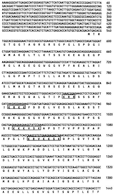
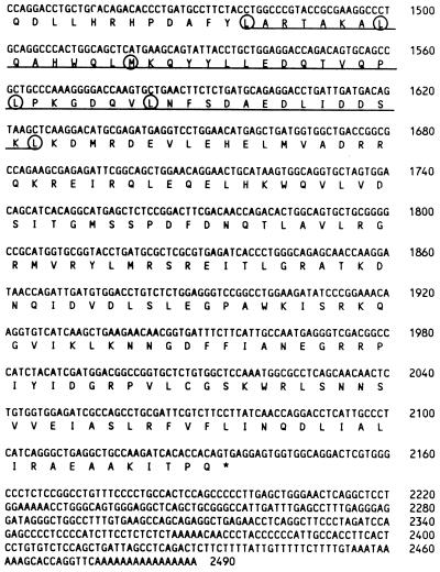
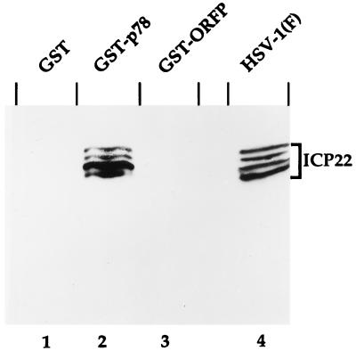
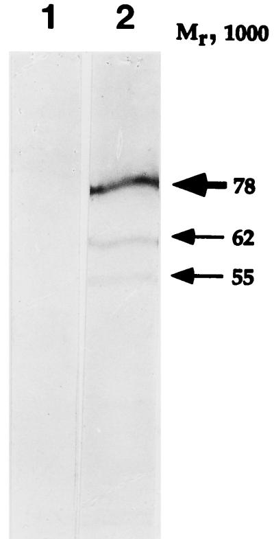
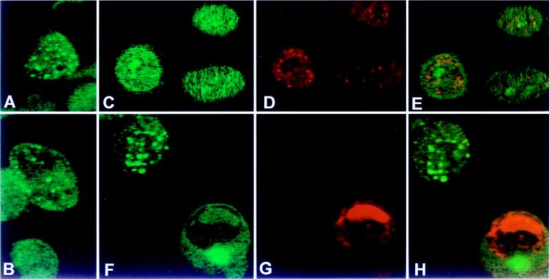
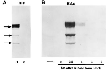
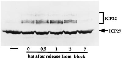
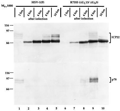
The herpes simplex virus 1 infected cell protein 22 (ICP22), the product of the alpha22 gene, is a nucleotidylylated and phosphorylated nuclear protein with properties of a transcriptional factor required for the expression of a subset of viral genes. Here, we report the following. (i) ICP22 interacts with a previously unknown cellular factor designated p78 in the yeast two-hybrid system. The p78 cDNA encodes a polypeptide with a distribution of leucines reminiscent of a leucine zipper. (ii) In uninfected and infected cells, antibody to p78 reacts with two major bands with an apparent Mr of 78,000 and two minor bands with apparent Mrs of 62, 000 and 55,000. (ii) p78 also interacts with ICP22 in vitro. (iii) In uninfected cells, p78 was dispersed largely in the nucleoplasm in HeLa cells and in the nucleoplasm and cytoplasm in HEp-2 cells. After infection, p78 formed large dense bodies which did not colocalize with the viral regulatory protein ICP0. (iv) Accumulation of p78 was cell cycle dependent, being highest very early in S phase. (v) The accumulation of ICP22 in synchronized cells was highest in early S phase, in contrast to the accumulation of another protein, ICP27, which was relatively independent of the cell cycle. (vi) In the course of the cell cycle, ICP22 was transiently modified in an aberrant fashion, and this modification coincided with expression of p78. The results suggest that ICP22 interacts with and may be stabilized by cell cycle-dependent proteins.


Figures

FIG. 1
FIG. 1
Nucleotide and amino acid sequences of p78. The amino acid sequence is shown below the DNA sequence. The underlined sequence refers to the putative leucine zipper region. Hydrophobic residues occurring every seven amino acids are circled. The boxed amino acid sequences are potential nuclear localization signals. Numbers on the right refer to the DNA sequence.
FIG. 1
FIG. 1
Nucleotide and amino acid sequences of p78. The amino acid sequence is shown below the DNA sequence. The underlined sequence refers to the putative leucine zipper region. Hydrophobic residues occurring every seven amino acids are circled. The boxed amino acid sequences are potential nuclear localization signals. Numbers on the right refer to the DNA sequence.
FIG. 2
FIG. 2
Photographic image of cell proteins bound to GST fusion proteins, electrophoretically separated in denaturing gels and reacted with a serum to ICP22. Recombinant pGEX vectors were grown in E. coli BL21, and fusion proteins were isolated as recommended by the manufacturer (Pharmacia). Fusion protein (2 to 5 μg) was mixed with HSV-1(F)-infected HeLa extract and incubated at 4°C for 2 to 4 h. Beads were collected by centrifugation, washed three times with PBS* (see Materials and Methods), and resuspended in 60 μl of 2× disruption buffer. Proteins were separated in 7% denaturing polyacrylamide gel, transferred to nitrocellulose, blocked, reacted with R77 and then with a goat anti-rabbit antibody conjugated to alkaline phosphatase, and processed as described by the manufacturer (Bio-Rad). Lanes 1 to 3, HeLa cell extract bound to GST, GST-p78, and GST-ORF P, respectively; lane 4, cell extract from infected HeLa cells.
FIG. 3
FIG. 3
Photographic image of HeLa cell proteins, electrophoretically separated in denaturing gels and reacted with a preimmune serum (lane 1) or with a serum to p78 (lane 2). Uninfected HeLa cell extract was separated in a 10% denaturing polyacrylamide gel, blocked, reacted with a rabbit preimmune serum or serum to p78 (see Materials and Methods) and then with a goat anti-rabbit antibody conjugated to alkaline phosphatase, and processed as described by the manufacturer (Bio-Rad). The major p78 band resolves as a doublet in lower-percentage polyacrylamide gels.
FIG. 4
FIG. 4
Photomicrographs of uninfected (A and B) and HSV-1(F)-infected (C to E and F to H) HeLa cells (B) and Hep-2 cells (A, C to E, and F to H) reacted with an antibodies to p78 and ICP0 (see Materials and Methods). Infected cells were fixed at 3 h after infection (C to E) or at 18 h after infection (F to H). Immunofluorescence was done as described in Materials and Methods. (A, B, C, and F) FITC-labeled anti-rabbit IgG antibody to p78 antiserum; (D and G) Texas red-labeled anti-mouse IgG to the ICP0 antibody; (E and H) overlays of panels C and D and panels F and G, respectively.
FIG. 5
FIG. 5
Photographic images of uninfected HFF and HeLa cell extracts, electrophoretically separated in denaturing gels and reacted with a serum to p78. (A) HFF extracts were prepared at 50% cell confluency (lane 1) and at 100% cell confluency (lane 2), separated in 7% denaturing gels, transferred to nitrocellulose, blocked, and reacted with a serum to p78 and then with a goat anti-rabbit antibody conjugated to alkaline phosphatase. Gel loading was monitored by staining for actin (not shown). (B) HeLa cell extracts were prepared from unsynchronized cells (leftmost lane) and from synchronized cells at 0, 0.5, 1, 3, and 7 h after release from block (see Materials and Methods) and processed as described above.
FIG. 6
FIG. 6
Photographic image of wild-type virus-infected HeLa cell extracts, separated in denaturing gels and reacted with sera to viral α proteins. Unsynchronized HeLa cells (leftmost lane) and synchronized HeLa cells were infected with HSV-1(F) at the time points shown after release from block (see Materials and Methods) for 24 h. Cell extracts were separated in 7% denaturing polyacrylamide gels, transferred to nitrocellulose sheets, blocked, reacted with sera to ICP22 and ICP27 and then with a goat anti-mouse (ICP27) or a goat anti-rabbit (for ICP22) antibody conjugated to alkaline phosphatase, and processed as recommended by the manufacturer.
FIG. 7
FIG. 7
Photographic image of cell extracts infected with wild-type virus or with recombinant R7353, separated in denaturing gels and reacted with sera to ICP22 and p78. Synchronized HeLa cells were infected at 0.5 h after release from block with either HSV-1(F) (lanes 1 to 5) or recombinant R7353 (lanes 6 to 10), and extracts were prepared at the times indicated after infection. Cell extracts were separated in duplicate 7% denaturing polyacrylamide gels, transferred to nitrocellulose sheets, and reacted with sera to ICP22 (upper panel) and p78 (lower panel).

References

    1. Ackermann M, Braun D K, Pereira L, Roizman B. Characterization of herpes simplex virus 1 proteins 0, 4, and 27 with monoclonal antibodies. J Virol. 1984;52:108–118. - PMC - PubMed
    1. Ackermann M, Sarmiento M, Roizman B. Application of antibody to synthetic peptides for characterization of the intact and truncated α22 protein specified by herpes simplex 1 and the R325 α22− deletion mutant. J Virol. 1985;56:207–215. - PMC - PubMed
    1. Altschul S F, Gish W, Miller W, Myers E W, Lipman D J. Basic local alignment search tool. J Mol Biol. 1990;215:403–410. - PubMed
    1. Baxevanis A D, Vinson C R. Interactions of coiled coils in transcription factors: where is the specificity? Curr Opin Genet Dev. 1993;3:278–285. - PubMed
    1. Blaho J A, Mitchell C, Roizman B. Guanylylation and adenylylation of the α regulatory proteins of the herpes simplex virus require a viral β or γ function. J Virol. 1993;67:3891–3900. - PMC - PubMed

Publication types

MeSH terms

Associated data