. 1998 Nov;72(11):8568-77.
doi: 10.1128/JVI.72.11.8568-8577.1998.

Circular intermediates of recombinant adeno-associated virus have defined structural characteristics responsible for long-term episomal persistence in muscle tissue

Affiliations

Circular intermediates of recombinant adeno-associated virus have defined structural characteristics responsible for long-term episomal persistence in muscle tissue

D Duan et al. J Virol. 1998 Nov.

Erratum in

  • J Virol 1999 Jan;73(1):861

Abstract

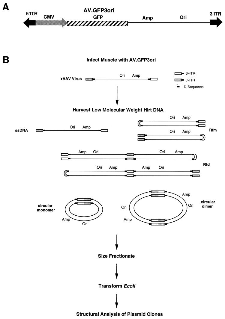
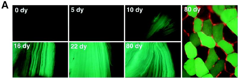
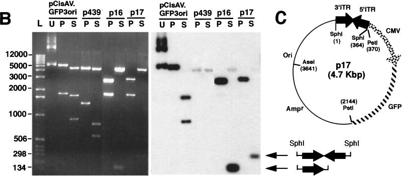
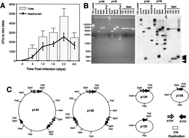
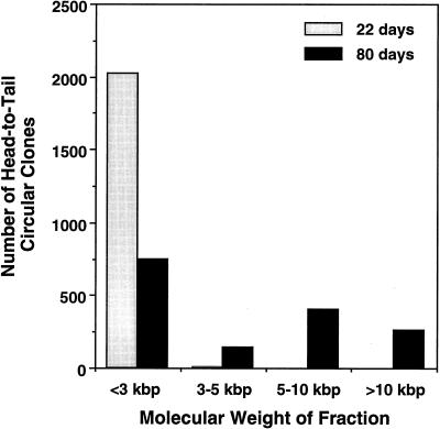
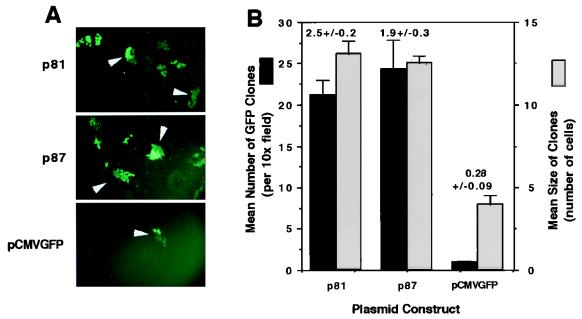
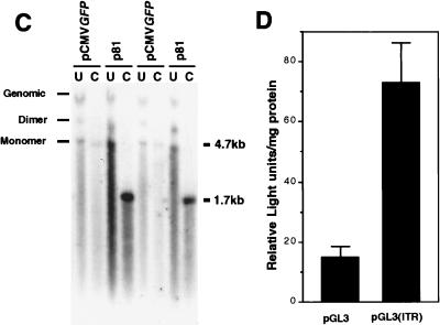
Adeno-associated viral (AAV) vectors have demonstrated great utility for long-term gene expression in muscle tissue. However, the mechanisms by which recombinant AAV (rAAV) genomes persist in muscle tissue remain unclear. Using a recombinant shuttle vector, we have demonstrated that circularized rAAV intermediates impart episomal persistence to rAAV genomes in muscle tissue. The majority of circular intermediates had a consistent head-to-tail configuration consisting of monomer genomes which slowly converted to large multimers of >12 kbp by 80 days postinfection. Importantly, long-term transgene expression was associated with prolonged (80-day) episomal persistence of these circular intermediates. Structural features of these circular intermediates responsible for increased persistence included a DNA element encompassing two viral inverted terminal repeats (ITRs) in a head-to-tail orientation, which confers a 10-fold increase in the stability of DNA following incorporation into plasmid-based vectors and transfection into HeLa cells. These studies suggest that certain structural characteristics of AAV circular intermediates may explain long-term episomal persistence with this vector. Such information may also aid in the development of nonviral gene delivery systems with increased efficiency.


Figures

FIG. 1
FIG. 1
Isolation of rAAV circular intermediates. With the aid of a rAAV cis-acting plasmid, pCisAV.GFP3ori, recombinant AAV virus (AV.GFP3ori) was generated (A). This vector carries a CMV promoter/enhancer, GFP transgene cassette, ampicillin resistance gene (Amp), and bacterial replication origin (Ori). (B) Strategy for isolation of rAAV circular intermediates. ssDNA, single-stranded DNA; Rfm, replication form monomer; Rfd, replication form dimer.
FIG. 2
FIG. 2
Formation of rAAV head-to-tail circular intermediates following in vivo transduction of muscle tissue. The tibialis anterior muscles of 4- to 5-week old C57BL/6 mice were infected with AV.GFP3ori (3 × 10¹⁰ particles) in HEPES-buffered saline (30 μl). GFP expression (A) was analyzed by direct immunofluorescence of freshly excised tissues and/or in formalin-fixed cryopreserved tissue sections in four independently injected muscle samples harvest at 0, 5, 10, 16, 22, and 80 days (dy) postinfection. GFP expression was detected at low levels beginning at 10 days and was maximum at 22 days postinfection. Expression remained stable to 80 days, at which time more than 50% of the tissue was positive (80-day tissue cross section counterstained with propidium iodide). Hirt DNA was isolated from muscle samples at each of the various time points was used to transform E. coli. Rescued plasmids (p439, p16, and p17; all isolated from muscle tissue at 22 days postinfection) were analyzed by Southern blotting (B; agarose gel [left] and ITR-probed blot [right]). Lanes U, uncut; P, PstI cut; S, SphI cut. Positions of molecular weight DNA standards (lane L) are given at the left in base pairs. (C) Schematic drawing of the most predominant type of head-to-tail circular AAV intermediate plasmids rescued from bacteria, showing the structure of p17 as an example (SphI site flanking the 3′ ITR designated the first base pair in the plasmid). Other typical clones included those with fewer than two ITRs as shown for p16. SphI digestion of p16 and p17 plasmids released ITR-hybridizing fragments of approximately 140 and 300 bp, respectively. The slightly lower than predicted apparent molecular size for these ITR fragments (364 bp for an ITR/ITR array) likely represents anomalous migration due to the high secondary structure of inverted repeats within ITRs. Additional restriction enzyme analyses used to determine these structures included double and single digests with SphI, PstI, AseI, and/or SmaI (data not shown). Although sequence analysis of p17 and p16 using nested primers to 5′ and 3′ ITRs also confirmed the ITR orientations shown, complete sequence through the central regions of inverted ITR arrays in p17 and similar structures was not possible due to the high secondary structure. Hence, at present it is impossible to rule out the possibility of small deletions in the central junctional regions of inverted ITRs. An example of an atypical clone (p439) rescued from bacteria with unknown structure is also shown.
FIG. 2
FIG. 2
Formation of rAAV head-to-tail circular intermediates following in vivo transduction of muscle tissue. The tibialis anterior muscles of 4- to 5-week old C57BL/6 mice were infected with AV.GFP3ori (3 × 10¹⁰ particles) in HEPES-buffered saline (30 μl). GFP expression (A) was analyzed by direct immunofluorescence of freshly excised tissues and/or in formalin-fixed cryopreserved tissue sections in four independently injected muscle samples harvest at 0, 5, 10, 16, 22, and 80 days (dy) postinfection. GFP expression was detected at low levels beginning at 10 days and was maximum at 22 days postinfection. Expression remained stable to 80 days, at which time more than 50% of the tissue was positive (80-day tissue cross section counterstained with propidium iodide). Hirt DNA was isolated from muscle samples at each of the various time points was used to transform E. coli. Rescued plasmids (p439, p16, and p17; all isolated from muscle tissue at 22 days postinfection) were analyzed by Southern blotting (B; agarose gel [left] and ITR-probed blot [right]). Lanes U, uncut; P, PstI cut; S, SphI cut. Positions of molecular weight DNA standards (lane L) are given at the left in base pairs. (C) Schematic drawing of the most predominant type of head-to-tail circular AAV intermediate plasmids rescued from bacteria, showing the structure of p17 as an example (SphI site flanking the 3′ ITR designated the first base pair in the plasmid). Other typical clones included those with fewer than two ITRs as shown for p16. SphI digestion of p16 and p17 plasmids released ITR-hybridizing fragments of approximately 140 and 300 bp, respectively. The slightly lower than predicted apparent molecular size for these ITR fragments (364 bp for an ITR/ITR array) likely represents anomalous migration due to the high secondary structure of inverted repeats within ITRs. Additional restriction enzyme analyses used to determine these structures included double and single digests with SphI, PstI, AseI, and/or SmaI (data not shown). Although sequence analysis of p17 and p16 using nested primers to 5′ and 3′ ITRs also confirmed the ITR orientations shown, complete sequence through the central regions of inverted ITR arrays in p17 and similar structures was not possible due to the high secondary structure. Hence, at present it is impossible to rule out the possibility of small deletions in the central junctional regions of inverted ITRs. An example of an atypical clone (p439) rescued from bacteria with unknown structure is also shown.
FIG. 3
FIG. 3
Frequency of circular intermediate formation in muscle tissue following transduction with rAAV. Hirt DNAs isolated from rAAV-infected tibialis muscle samples were used to transform E. coli, and the rescued plasmids were analyzed by Southern blotting as previous described (more than 20 clones were analyzed from at least two independent muscle samples for each time point). (A) Numbers (mean ± standard error) of total head-to-tail circular intermediate clones (line) and total ampicillin-resistant bacteria clones (bar) isolated from each tibialis anterior muscle at 0, 5, 10, 16, 22, and 80 days postinfection. Only plasmids which contained one to two ITRs were included in the estimation of total head-to-tail circular intermediates. Plasmids which demonstrated an absence of ITR-hybridizing SphI fragments (between 150 to 300 bp) were omitted from the calculations. (B) Diversity of ITR arrays found in head-to-tail circular intermediates at 80 days postinfection. The Southern blot was probed with ITR sequences and represents circular intermediates with one to three ITRs. SphI fragments which excise the inverted ITR arrays and hybridize to ITR probes are marked by arrowheads at the right. Sizes of molecular weight standards are indicated at the left in base pairs. Additional restriction enzyme analysis was used to determine the structure of monomer and multimer circular intermediates. Examples are shown for two multimer circular intermediates (p136 and p143) which contain approximately three AAV genomes. Undigested p136 and p143 plasmids migrate at >12 kbp whereas the predominant forms of head-to-tail undigested circular intermediates at 22 days migrate at 2.5 kbp (data not shown). The digestion pattern of p136 is consistent with a uniform head-to-tail configuration of three genomes and is indistinguishable from the digestion pattern of p139, which contains one circularized genome (undigested p139 migrates at 2.5 kbp [data not shown; also see results for p17 in Fig. 2]). In contrast, p136 exhibits a more complex head-to-tail multimer circular intermediate which has various deletions and duplications within the ITR arrays. (C) Predicted structures of five representative intermediates (complete data for structural analysis are not given).
FIG. 4
FIG. 4
Molecular sizes of native unamplified circular intermediates in muscle tissue. Hirt DNA from AV.GFP3ori-infected muscle was size fractionated by electrophoresis, and fractions of various molecular weights were transformed into E. coli. Results demonstrate the abundance of head-to-tail circular intermediates at each of the given molecular sizes at 22 and 80 days after infection with the rAAV shuttle vector. Fractions were sized according to the migration of linear double-stranded DNA standards under identical gel running conditions. The structures of circular intermediates were confirmed by Southern blot restriction analysis (data not shown).
FIG. 5
FIG. 5
Head-to-tail circular intermediates demonstrate increased stability of GFP expression following transient transfection in HeLa cells. Subconfluent monolayers of HeLa cells were cotransfected with p81, p87, or pCMVGFP and pRSVlacZ as an internal control for transfection efficiency as described in Materials and Methods. (A) Expansion of GFP clones after one passage (arrowheads). (B) Quantification of clone size (mean raw values) and clone numbers (normalized for transfection efficiency as determined by X-Gal staining for pRSVlacZ). Numbers above the bars represent quantification of GFP clones after passage 2 (also normalized for transfection efficiency). Results indicate the means (± standard errors) of duplicate experiments, with more than 20 fields quantified for each experimental point. The persistence of transfected p81 and pCMVGFP plasmid DNA at passage 7 posttransfection was evaluated by genomic Southern blotting of total cellular DNA hybridized against a 32P-labeled GFP probe (C; results from two independent transfections are shown). In these studies, control cultures cotransfected with pRSVlacZ demonstrated a less than 25% variation in transfection efficiency, as determined by X-Gal staining of cells at 48 h posttransfection. Lanes: U, uncut; C, PstI cut. The migration of uncut dimer and monomer plasmids forms are marked on the left. PstI digestion of the plasmids results in bands at 4.7 kb (pCMVGFP; single PstI site in plasmid) and 1.7 kb (p81; two PstI sites flanking the GFP gene). The band marked Genomic in undigested lanes was also seen with DNA from untransfected cells (data not shown) and hence is nonspecific hybridization of probe to the high concentration of DNA in this area of the gel. To determine whether the head-to-tail ITR array within circular intermediates was responsible for increases in the persistence of GFP expression, the head-to-tail ITR DNA element was subcloned into the luciferase plasmid pGL3 to generate pGL3(ITR). (D) Luciferase transgene expression following transfection with pGL3 and pGL3(ITR) at 10 days (passage 2) posttransfection. Results are the means (± standard errors) for triplicate experiments and are normalized for transfection efficiency by using a dual renilla-luciferase reporter vector (pRLSV40; Promega).
FIG. 5
FIG. 5
Head-to-tail circular intermediates demonstrate increased stability of GFP expression following transient transfection in HeLa cells. Subconfluent monolayers of HeLa cells were cotransfected with p81, p87, or pCMVGFP and pRSVlacZ as an internal control for transfection efficiency as described in Materials and Methods. (A) Expansion of GFP clones after one passage (arrowheads). (B) Quantification of clone size (mean raw values) and clone numbers (normalized for transfection efficiency as determined by X-Gal staining for pRSVlacZ). Numbers above the bars represent quantification of GFP clones after passage 2 (also normalized for transfection efficiency). Results indicate the means (± standard errors) of duplicate experiments, with more than 20 fields quantified for each experimental point. The persistence of transfected p81 and pCMVGFP plasmid DNA at passage 7 posttransfection was evaluated by genomic Southern blotting of total cellular DNA hybridized against a 32P-labeled GFP probe (C; results from two independent transfections are shown). In these studies, control cultures cotransfected with pRSVlacZ demonstrated a less than 25% variation in transfection efficiency, as determined by X-Gal staining of cells at 48 h posttransfection. Lanes: U, uncut; C, PstI cut. The migration of uncut dimer and monomer plasmids forms are marked on the left. PstI digestion of the plasmids results in bands at 4.7 kb (pCMVGFP; single PstI site in plasmid) and 1.7 kb (p81; two PstI sites flanking the GFP gene). The band marked Genomic in undigested lanes was also seen with DNA from untransfected cells (data not shown) and hence is nonspecific hybridization of probe to the high concentration of DNA in this area of the gel. To determine whether the head-to-tail ITR array within circular intermediates was responsible for increases in the persistence of GFP expression, the head-to-tail ITR DNA element was subcloned into the luciferase plasmid pGL3 to generate pGL3(ITR). (D) Luciferase transgene expression following transfection with pGL3 and pGL3(ITR) at 10 days (passage 2) posttransfection. Results are the means (± standard errors) for triplicate experiments and are normalized for transfection efficiency by using a dual renilla-luciferase reporter vector (pRLSV40; Promega).

References

    1. Afione S A, Conrad C K, Kearns W G, Chunduru S, Adams R, Reynolds T C, Guggino W B, Cutting G R, Carter B J, Flotte T R. In vivo model of adeno-associated virus vector persistence and rescue. J Virol. 1996;70:3235–3241. - PMC - PubMed
    1. Beaton A, Palumbo P, Berns K I. Expression from the adeno-associated virus p5 and p19 promoters is negatively regulated in trans by the Rep protein. J Virol. 1989;63:4450–4454. - PMC - PubMed
    1. Bennett J, Duan D, Engelhardt J F, Maguire A M. Real-time, noninvasive in vivo assessment of adeno-associated virus-mediated retinal transduction. Investig Ophthalmol Visual Sci. 1997;38:2857–2863. - PubMed
    1. Cheung A K, Hoggan M D, Hauswirth W W, Berns K I. Integration of the adeno-associated virus genome into cellular DNA in latently infected human Detroit 6 cells. J Virol. 1980;33:739–748. - PMC - PubMed
    1. Clark K R, Sferra T J, Johnson P R. Recombinant adeno-associated viral vectors mediate long-term transgene expression in muscle. Hum Gene Ther. 1997;8:659–669. - PubMed

Publication types

MeSH terms

LinkOut - more resources