. 1998 Nov;72(11):8578-85.
doi: 10.1128/JVI.72.11.8578-8585.1998.

Intracellular localization of poliovirus plus- and minus-strand RNA visualized by strand-specific fluorescent In situ hybridization

Affiliations

Intracellular localization of poliovirus plus- and minus-strand RNA visualized by strand-specific fluorescent In situ hybridization

R Bolten et al. J Virol. 1998 Nov.

Abstract

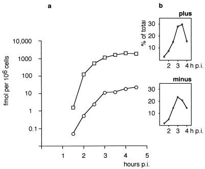
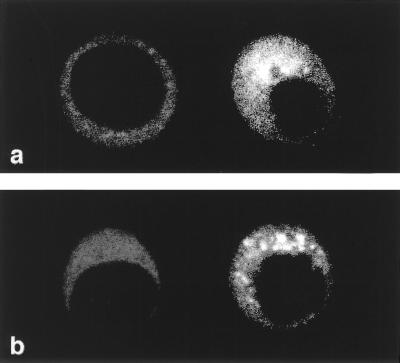
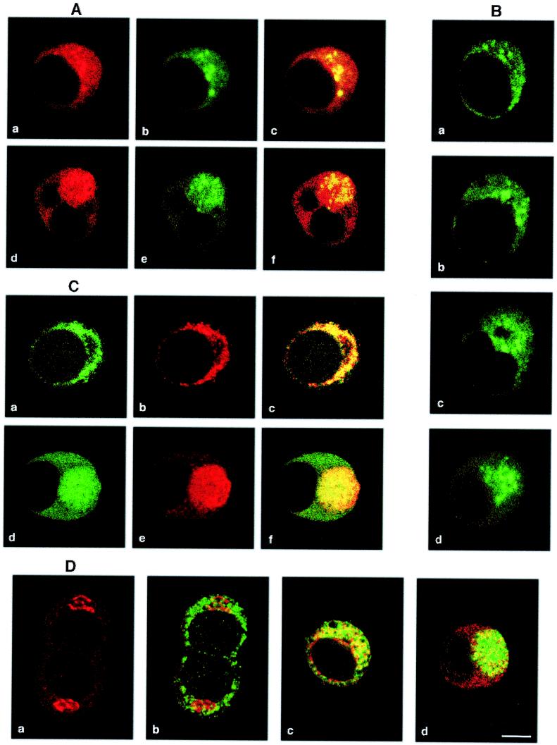
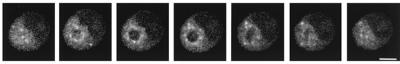
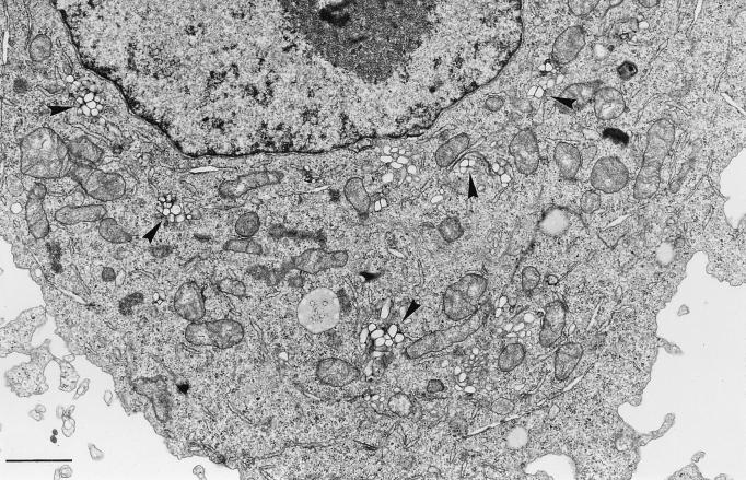
The time courses of poliovirus plus- and minus-strand RNA synthesis in infected HEp-2 cells were monitored separately, using a quantitative RNase assay. In parallel, viral RNA and proteins were located in situ by confocal microscopy within cells fixed by a protocol determined to retain their native size and shape. Plus- and minus-strand RNAs were visualized by fluorescent in situ hybridization (FISH) with strand-specific riboprobes. The probes were labelled with different fluorochromes to allow for the simultaneous detection of plus- and minus-strand RNA. The FISH experiments showed minus-strand RNA to be present in distinct, regularly sized, round structures throughout the viral replication cycle. Plus-strand RNA was found in the same structures and also in smaller clusters of vesicles. Association of viral RNA with membranes was demonstrated by combining FISH with immunofluorescence (IF) detection of the viral 2B- and 2C-containing P2 proteins, which are known to be markers for virus-induced membranes. At early times postinfection, the virus-induced membranous structures were distributed through most of the cytoplasm, whereas around peak RNA synthesis, both RNA-associated membranous structures migrated to the center of the cell. During this process, the plus- and minus-strand-containing larger structures stayed as recognizable entities, whereas the plus-strand-containing granules coalesced into a juxtanuclear area of membranous vesicles. An involvement of Golgi-derived membranes in the formation of virus-induced vesicles and RNA synthesis early in infection was investigated by IF with 2C- and Golgi-specific antibodies.


Figures

FIG. 1
FIG. 1
Kinetics of plus- and minus-strand RNA synthesis in PV-infected HEp-2 cells as determined by RPA. (a) Time p.i. versus amounts (logarithmic scale) of plus (□)- and minus (○)-strand RNA synthesized. (b) Peak synthesis of plus- and minus-strand RNA is between 3 and 3.5 h p.i.
FIG. 2
FIG. 2
Evaluation of background staining in FISH with uninfected (left) and PV-infected (right) cells in the same microscopic field with a Texas red-labelled plus-strand-specific probe (a) and an FITC-labelled minus-strand-specific probe (b). For an explanation of structural details, see the legend to Fig. 3.
FIG. 3
FIG. 3
Viral RNA and protein detected in PV-infected HEp-2 cells by fluorescent confocal microscopy. Bar, 5 μm. (A) Simultaneous visualization of viral plus- and minus-strand RNA in the same cell by double-labelling FISH. Plus-strand RNA (panels a and d), detected with a Texas red-labelled riboprobe of minus polarity, is found in several small granules, at first dispersed and later in infection concentrated in a juxtanuclear area. Minus-strand RNA (panels b and e), detected with an FITC-labelled riboprobe of plus polarity, is present in larger, distinct granules. Superimposed plus- and minus-strand detection shows colocalization of both signals in yellow (panels c and f). Panels a to c, 2.5 h p.i.; panels d to f, 3.5 h p.i. (B) Minus-strand RNA, detected with an FITC-labelled riboprobe as for panel A, remains in distinct granules throughout the replication cycle. Panel a, 2.25 h p.i.; panel b, 2.5 h p.i.; panel c, 3.0 h p.i.; panel d, 3.5 h p.i. (C) Simultaneous visualization of viral plus-strand RNA and viral protein 2C by combined FISH and IF. Plus-strand RNA (panels a and d) is detected with an FITC-labelled riboprobe. Protein 2C and 2C-containing precursors (panels b and e) are detected with an anti-2C MAb and Texas red-labelled anti-mouse IgG. Superimposed FISH and IF show that early in infection, plus-strand RNA and 2C are colocalized (yellow in panel c); at later times some dissociation of 2C from RNA is visible (panel f). Panels a to c, 2.0 h p.i.; panels d to f, 3.5 h p.i. (D) IF with a MAb against Golgi protein p58 detected with Texas red-labelled anti-mouse IgG, combined with IF with a polyclonal rabbit anti-2C Ab detected with an FITC-labelled anti-rabbit IgG. Panel a, intact Golgi complexes at 2 h p.i. Panel b, the same cells as in panel a, showing IF with anti-2C Ab superimposed. Little colocalization (yellow) of the two targets is found. Panel c, superimposed pictures of a cell double labelled by IF with anti-p58 and anti-2C Ab at 2.5 h p.i. The Golgi marker and viral protein 2C colocalize partially. Panel d, superimposed pictures as in panels b and c at 3 h p.i. The Golgi marker is distributed in the cytoplasm and is colocalized with protein 2C mainly at the border of the juxtanuclear area of vesicles.
FIG. 4
FIG. 4
FISH for minus-strand detection in a series of optical sections, spaced 0.4 μm apart, through an infected cell at 2.75 h p.i. Minus-strand RNA is found in single spherical granules of approximately 0.5 μm in diameter in a symmetrical array around an unlabelled center. Bar, 5 μm.
FIG. 5
FIG. 5
Electron micrograph of a PV-infected HEp-2 cell at 2 h p.i., showing individual small clusters of virus-induced vesicles (arrowheads). Bar, 1 μm.

References

    1. Aldabe R, Barco A, Carrasco L. Membrane permeabilization by poliovirus proteins 2B and 2BC. J Biol Chem. 1996;271:23134–23137. - PubMed
    1. Aldabe R, Carrasco L. Induction of membrane proliferation by poliovirus proteins 2C and 2BC. Biochem Biophys Res Commun. 1995;206:64–76. - PubMed
    1. Andino R, Rieckhof G E, Baltimore D. A functional ribonucleoprotein complex forms around the 5′ end of poliovirus RNA. Cell. 1990;63:369–380. - PubMed
    1. Banerjee R, Echeverri A, Dasgupta A. Poliovirus-encoded 2C polypeptide specifically binds to the 3′-terminal sequences of viral negative-strand RNA. J Virol. 1997;71:9570–9578. - PMC - PubMed
    1. Baron M H, Baltimore D. In vitro copying of viral positive strand RNA by poliovirus replicase. Characterization of the reaction and its products. J Biol Chem. 1982;257:12359–12366. - PubMed

Publication types

LinkOut - more resources