. 1998 Nov;72(11):8738-46.
doi: 10.1128/JVI.72.11.8738-8746.1998.

Specific encapsidation of nodavirus RNAs is mediated through the C terminus of capsid precursor protein alpha

Affiliations

Specific encapsidation of nodavirus RNAs is mediated through the C terminus of capsid precursor protein alpha

A Schneemann et al. J Virol. 1998 Nov.

Abstract

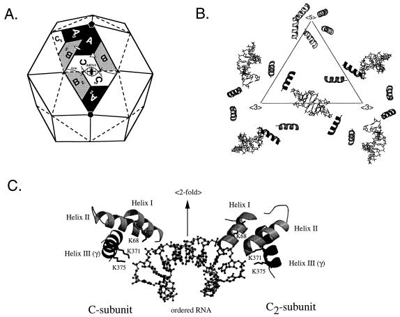
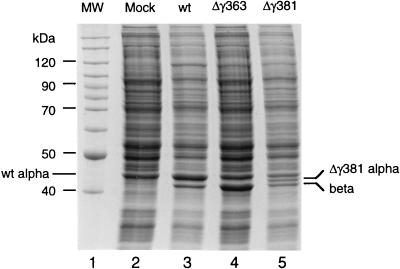
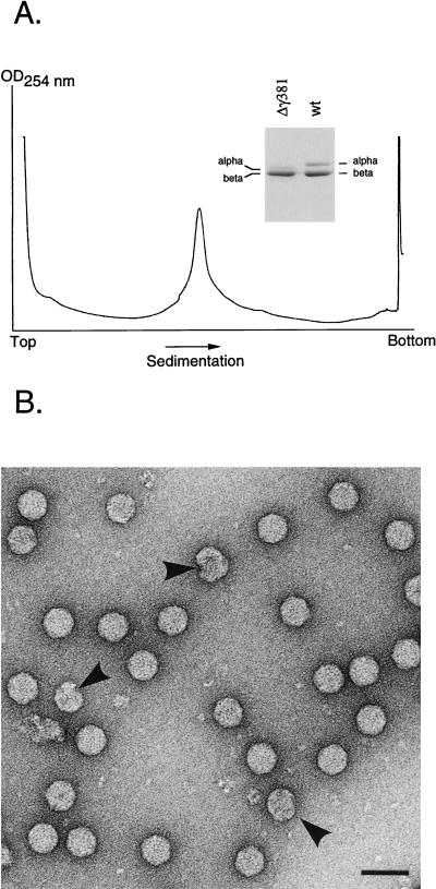
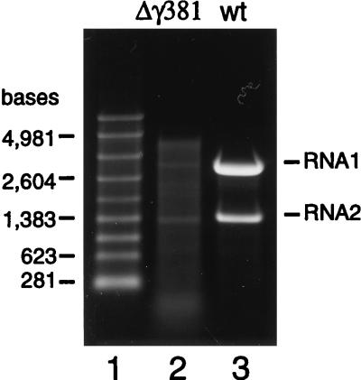
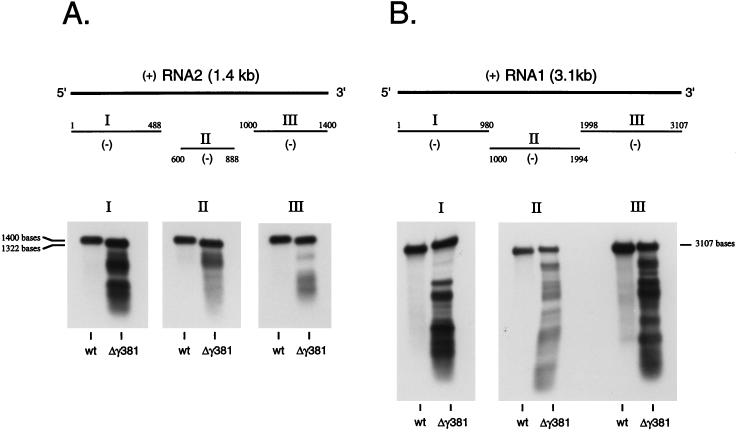
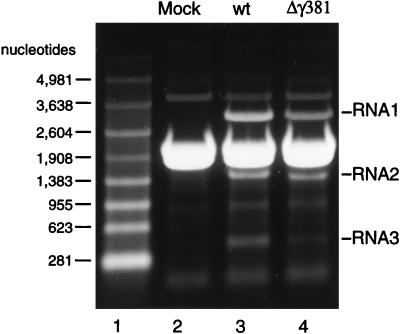
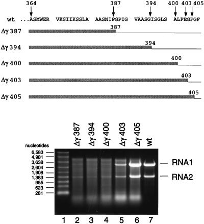
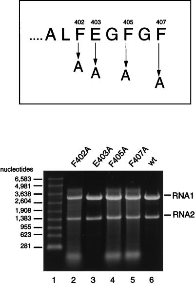
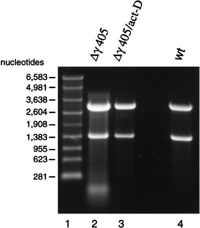
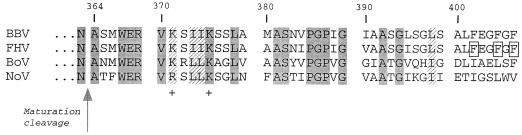
Flock house virus (FHV) is a small icosahedral insect virus with a bipartite, messenger-sense RNA genome. Its T=3 icosahedral capsid is initially assembled from 180 subunits of a single type of coat protein, capsid precursor protein alpha (407 amino acids). Following assembly, the precursor particles undergo a maturation step in which the alpha subunits autocatalytically cleave between Asn363 and Ala364. This cleavage generates mature coat proteins beta (363 residues) and gamma (44 residues) and is required for acquisition of virion infectivity. The X-ray structure of mature FHV shows that gamma peptides located at the fivefold axes of the virion form a pentameric helical bundle, and it has been suggested that this bundle plays a role in release of viral RNA during FHV uncoating. To provide experimental support for this hypothesis, we generated mutant coat proteins that carried deletions in the gamma region of precursor protein alpha. Surprisingly, we found that these mutations interfered with specific recognition and packaging of viral RNA during assembly. The resulting particles contained large amounts of cellular RNAs and varying amounts of the viral RNAs. Single-site amino acid substitution mutants showed that three phenylalanines located at positions 402, 405, and 407 of coat precursor protein alpha were critically important for specific recognition of the FHV genome. Thus, in addition to its hypothesized role in uncoating and RNA delivery, the C-terminal region of coat protein alpha plays a significant role in recognition of FHV RNA during assembly. A possible link between these two functions is discussed.


Figures

FIG. 1
FIG. 1
Organization of gamma peptides in the FHV particle and their interaction with genomic RNA. (A) Schematic representation of the FHV capsid as a rhombic triacontahedron. Each trapezoid represents a protein subunit which initially consists of 407 amino acids. The labels A, B, and C represent the three subunits in each of the 60 icosahedral asymmetric units in the T=3 particle. Although A, B, and C represent identical gene products, they are not related by strict symmetry and they are structurally slightly different. In mature virions, most subunits are present as proteins beta and gamma, which are generated by autocatalytic cleavage of the precursor protein between residues Asn363 and Ala364. Note that ordered genomic RNA is visible at the icosahedral twofold axes (shown as a solid oval) of the virion. (B) Organization of the internally located gamma peptides and the ordered genomic RNA as seen in the X-ray structure. The triangle represents the central asymmetric unit shown in panel A. The first 18 residues of gamma form an amphipathic alpha helix, while the remaining 26 residues are not visible. Gamma peptides associated with the A subunits form a pentameric helical bundle at the fivefold axes of the virion. This helical bundle has been hypothesized to play a role in FHV uncoating. Gamma peptides associated with the C subunits contact the genomic RNA (shown as stick diagrams), which forms double-helical segments at the icosahedral twofold axes of the virion. (C) Close-up view of the interaction of the gamma peptides with genomic RNA (shown as a ball and stick model). The view is perpendicular to the icosahedral twofold axis, showing specific interactions between the phosphodiester backbone of FHV RNA and the side chains of lysine residues 68, 371, and 375. Lys68 is located on helix I, which consists of residues 61 to 73 of protein beta. Lys371 and Lys375 are located on helix III, which is part of the gamma peptide. Helix II, formed by residues 341 and 353, does not interact with the encapsidated RNA. All interactions are with the C subunit and its twofold related partner (C2 in the diagram shown in panel A). For clarity, only the helical domains of the coat protein subunits are shown.
FIG. 2
FIG. 2
Gel electrophoresis of lysates prepared from Drosophila cells transfected with wt FHV RNA1 and wt or mutant FHV transcript RNA2. Cell monolayers containing 107 Drosophila cells were transfected with a mixture of approximately 100 ng of RNA1 and 100 ng of RNA2, as described in Materials and Methods. At 24 h after transfection, cells were dislodged into the growth medium, collected by centrifugation, and washed twice with 0.5 ml of phosphate-buffered saline (PBS). The final cell pellet was resuspended in 100 μl of PBS, mixed with an equal volume of 2× electrophoresis buffer, and heated to 95°C for 10 min. Aliquots of 10 μl (approximately 5 × 10⁵ cells) were electrophoresed through an SDS–12% polyacrylamide gel, followed by staining with Coomassie brilliant blue. Gamma peptide migrated off the gel under the conditions used. Lane 1, molecular weight markers; lane 2, lysate from mock-transfected cells; lane 3, lysate from cells transfected with wt RNA1 and wt RNA2 (note that wt protein alpha comigrates with a cellular protein, probably actin); lane 4, lysate from cells transfected with wt RNA1 and Δγ363 RNA2; lane 5, lysate from cells transfected with wt RNA1 and Δγ381 RNA2.
FIG. 3
FIG. 3
Sucrose gradient sedimentation profile and electron microscopy of Δγ381 particles. (A) Monolayers of Drosophila cells (approximately 1.2 × 10⁸ cells) were transfected with wt RNA1 and Δγ381 RNA2, as described in Materials and Methods. After 24 h, cells were lysed and virus particles were pelleted through a 30% (wt/wt) sucrose cushion. The resuspended pellet was layered on a linear 10 to 40% (wt/wt) sucrose gradient and centrifuged at 274,000 × g for 1.5 h at 4°C. The gradient was fractionated with continuous absorbance at 254 nm. OD, optical density. (Inset) Electrophoretic analysis of proteins in the peak fraction and comparison with wt FHV. Gamma peptide migrated off the gel under the conditions used. Proteins were stained with Coomassie brilliant blue. (B) Electron micrograph of negatively stained, gradient-purified Δγ381 particles. Arrows indicate aberrant structures. Bar, 100 nm.
FIG. 4
FIG. 4
Gel electrophoretic analysis of RNAs extracted from Δγ381 and wt FHV particles. RNA was extracted with phenol-chloroform from gradient-purified virions and electrophoresed through a denaturing 1% agarose-formaldehyde gel. Nucleic acids were visualized by staining with ethidium bromide. Lane 1, RNA size markers; lane 2, RNA extracted from Δγ381 particles; lane 3, RNA extracted from wt particles.
FIG. 5
FIG. 5
Northern blot analysis of RNA extracted from Δγ381 particles and wt FHV particles. RNA (100 ng of Δγ381 RNA per lane; 5 ng of wt RNA per lane) was size fractionated on a 1% agarose-formaldehyde gel and transferred to a nylon membrane. Following UV cross-linking, the immobilized RNAs were hybridized to digoxigenin-UTP-labeled negative-sense RNA probes complementary to three different regions of RNA2 or RNA1. Hybridization complexes were visualized by chemiluminescence. (A) Hybridization with probes complementary to RNA2. Probe I was complementary to nt 1 to 488, probe II was complementary to nt 600 to 888, and probe III was complementary to nt 1000 to 1400. Note that full-length RNA in the Δγ381 sample migrates slightly faster than full-length wt RNA2 due to a deletion of 78 nt. (B) Hybridization with probes complementary to RNA1. Probe I was complementary to nt 1 to 980, probe II was complementary to nt 1000 to 1994, and probe III was complementary to nt 1998 to 3107. Note that samples hybridized with probe I migrated at a slight angle during gel electrophoresis, explaining the apparently larger molecular size of RNA1 in the Δγ381 sample than in the wt sample.
FIG. 6
FIG. 6
Electrophoretic analysis of total cellular RNA isolated from Drosophila cells transfected either with wt FHV RNAs or RNAs purified from Δγ381 particles. Drosophila cell monolayers (107 cells) were transfected either with 100 ng of wt RNA1 and 100 ng of capped in vitro-synthesized wt RNA2 or with approximately 1.5 μg of total RNA purified from Δγ381 particles. After 24 h, total cellular RNA was purified from the transfected cells and an aliquot of 3 μg was size fractionated on a 1% agarose-formaldehyde gel. RNAs were visualized with ethidium bromide. Lane 1, RNA molecular size markers; lane 2, total RNA from mock-transfected cells; lane 3, total RNA from cells transfected with wt FHV RNAs; lane 4, total RNA from cells transfected with RNA purified from Δγ381 particles. Note that RNA2 in this lane migrated slightly faster than wt RNA2 in lane 3 due to a deletion of 78 nt. The major band between RNA1 and RNA2 represents the two rRNAs which did not resolve under the electrophoresis conditions used here.
FIG. 7
FIG. 7
Electrophoretic analysis of RNA packaged by five different coat protein deletion mutants. (Top) Amino acid sequence of the wt gamma peptide (residues 364 to 407) and schematic representation of the mutant coat proteins. Thin lines indicate deleted areas, whereas shaded boxes represent maintained residues. Numbers refer to terminal residues in the mutant coat proteins. (Bottom) Denaturing agarose gel showing RNAs extracted from gradient-purified mutant particles. Particles were synthesized and purified as described in Materials and Methods, and RNA was extracted with phenol-chloroform. Aliquots were size fractionated on a denaturing 1% agarose-formaldehyde gel, and nucleic acids were visualized by staining with ethidium bromide. Lane 1, RNA molecular size markers; lane 2, RNA extracted from Δγ387 particles; lane 3, RNA extracted from Δγ394 particles; lane 4, RNA extracted from Δγ400 particles; lane 5, RNA extracted from Δγ403 particles; lane 6, RNA extracted from Δγ405 particles; lane 7, RNA extracted from wt particles.
FIG. 8
FIG. 8
Electrophoretic analysis of RNA packaged by four different single amino acid substitution mutants. (Top) Sequence of the last eight amino acids of the gamma peptide and positions that were changed to alanine. (Bottom) Denaturing agarose gel showing RNAs extracted from gradient-purified particles. Particles were synthesized and purified as described in Materials and Methods, and RNA was extracted with phenol-chloroform. Aliquots were size fractionated on a denaturing 1% agarose-formaldehyde gel, and nucleic acids were visualized by staining with ethidium bromide. Lane 1, RNA molecular size markers; lane 2, RNA extracted from F402A particles; lane 3, RNA extracted from E403A particles; lane 4, RNA extracted from F405A particles; lane 5, RNA extracted from F407A particles; lane 6, RNA extracted from wt particles.
FIG. 9
FIG. 9
Effect of actinomycin D on RNA encapsidation by Δγ405 coat protein. Monolayers of Drosophila cells (approximately 1.2 × 10⁸ cells) were transfected with wt RNA1 and in vitro-synthesized, capped Δγ405 RNA, as described in Materials and Methods. At 4 h after transfection, actinomycin D was added to the culture medium at a final concentration of 5 μg/ml, and incubation was continued for 20 h. Particles were then gradient purified, and RNA was extracted with phenol and chloroform. An aliquot was analyzed on a denaturing 1% agarose-formaldehyde gel followed by staining with ethidium bromide. Lane 1, RNA molecular size markers; lane 2, RNA extracted from Δγ405 particles grown in the absence of actinomycin D; lane 3, RNA extracted from Δγ405 particles grown in the presence of actinomycin D; lane 4, RNA extracted from wt FHV particles.
FIG. 10
FIG. 10
Multiple alignment of amino acid sequences of gamma peptides from four insect nodaviruses: BBV, FHV, Boolarra virus (BoV), and NoV. Arrow indicates site of maturation cleavage. Shaded residues are identical in all viruses, whereas hatched residues are conserved. In BBV, FHV, and NoV, whose structures are known at high resolution, gamma peptides located at position C on the icosahedral surface lattice contact the phosphodiester backbone of encapsidated RNA via positively charged side chains at positions 371 and 375. Boxed phenylalanine residues at positions 402, 405, and 407 of FHV are required for specific encapsidation of the viral RNAs by coat precursor protein alpha.

References

    1. Ball L A. Requirements for the self-directed replication of flock house virus RNA 1. J Virol. 1995;69:720–727. . (Erratum, 69:2722.) - PMC - PubMed
    1. Cheng R H, Reddy V S, Olson N H, Fisher A J, Baker T S, Johnson J E. Functional implications of quasi-equivalence in a T=3 icosahedral animal virus established by cryo-electron microscopy and X-ray crystallography. Structure. 1994;2:271–282. - PMC - PubMed
    1. Convery M A, Rowsell S, Stonehouse N J, Ellington A D, Murray H I J B, Peabody D S, Phillips S E V, Stockley P G. Crystal structure of an aptamer-protein complex at 2.8 A resolution. Nat Struct Biol. 1998;5:133–139. - PubMed
    1. Dasmahapatra B, Dasgupta R, Ghosh A, Kaesberg P. Structure of the black beetle virus genome and its functional implications. J Mol Biol. 1985;182:183–189. - PMC - PubMed
    1. Dong X F, Natarajan P, Tihova M, Johnson J E, Schneemann A. Particle polymorphism caused by deletion of a peptide molecular switch in a quasi-equivalent virus. J Virol. 1998;72:6024–6033. - PMC - PubMed

Publication types

MeSH terms

LinkOut - more resources