. 1998 Nov;72(11):8797-805.
doi: 10.1128/JVI.72.11.8797-8805.1998.

CD4 promoter transactivation by human herpesvirus 6

Affiliations

CD4 promoter transactivation by human herpesvirus 6

L Flamand et al. J Virol. 1998 Nov.

Abstract

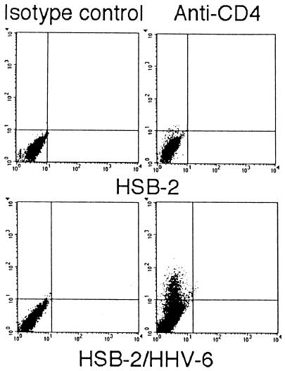
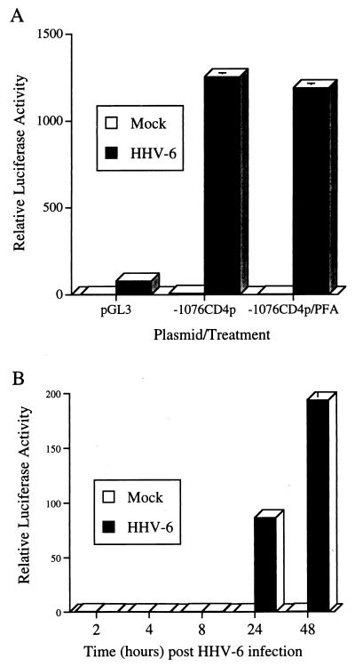
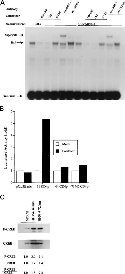
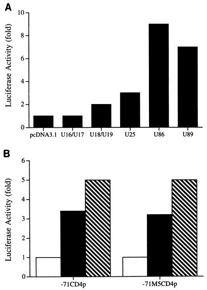
The observation that human herpesvirus 6 (HHV-6) can induce CD4 gene transcription and expression in CD4(-) cells was reported several years ago (P. Lusso, A. De Maria, M. Malnati, F. Lori, S. E. DeRocco, M. Baseler, and R. C. Gallo, Nature 349:533-535, 1991) and subsequently confirmed (P. Lusso, M. S. Malnati, A. Garzino-Demo, R. W. Crowley, E. O. Long, and R. C. Gallo, Nature 362:458-462, 1993; G. Furlini, M. Vignoli, E. Ramazzotti, M. C. Re, G. Visani, and M. LaPlaca, Blood 87:4737-4745, 1996). Our objective was to identify the mechanisms underlying such phenomena. Using reporter gene constructs driven by the CD4 promoter, we report that HHV-6 can efficiently transactivate such genetic elements. Activation of the CD4 promoter occurs in the presence of the viral DNA polymerase inhibitor phosphonoformic acid, which limits expression to the immediate-early and early classes of viral genes. Using deletion mutants and specific CD4 promoter mutants, we identified an ATF/CRE binding site located at nucleotides -67 to -60 upstream of the CD4 gene transcription start site that is important for HHV-6 transactivation. The ATF/CRE site is also essential for CD4 promoter activation by forskolin, an activator of adenylate cyclase. Using electrophoretic mobility shift assays and specific antibodies, we showed that CREB-1 binds specifically to the -79 to -52 region of the CD4 promoter. Last, we have identified two open reading frames (ORFs) of HHV-6, U86 and U89 from the immediate-early locus A, that can transactivate the CD4 promoter in HeLa cells. However, transactivation of the CD4 promoter by ORFs U86 and U89 is independent of the CRE element, suggesting that additional HHV-6 ORFs are likely to contribute to CD4 gene activation. Taken together, our results will help to understand the complex interactions occurring between HHV-6 and the CD4 promoter and provide additional information regarding the class of transcription factors involved in the control of CD4 gene expression.


Figures

FIG. 1
FIG. 1
Cell surface expression of CD4 after infection of HSB-2 cells with HHV-6. HSB-2 cells (CD4) were either treated with mock-infected culture fluid or infected with HHV-6 (GS strain). When signs of cytopathic effect were observed (5 to 7 days), cells were analyzed for CD4 surface expression by flow cytometry using PE-labeled anti-CD4 or isotype-matched control antibody. Top panels represent uninfected cells, while bottom panels depict HHV-6-infected cells.
FIG. 2
FIG. 2
(A) Activation of the CD4 promoter by HHV-6. HSB-2 cells were transfected with control (pGL3) or with −1076CD4p constructs and infected with HHV-6 or treated with mock-infected culture supernatant. PFA was added 1 h prior to infection, and PFA was kept throughout the experiment. Cells were harvested and lysed at 48 h postinfection, and luciferase activity was determined. Results (mean ± standard deviation), expressed as relative luciferase activity, are calculated from triplicate cultures and are representative of three experiments. (B) Kinetics of CD4 promoter activation by HHV-6. HSB-2 cells were transfected with the −1076CD4p construct and treated, the next day, with either mock-infected fluid or with HHV-6. At various times after infection, cell were harvested and lysed and luciferase activity was determined. Results (mean ± standard deviation) are calculated from triplicate cultures and are representative of three experiments.
FIG. 3
FIG. 3
(A) Schematic representation of wild-type (−1076CD4p) and mutant CD4 promoter luciferase reporter gene constructs. The cloning plasmid used for all of these constructs was the pGL3basic vector. The vertical line in constructs −1076M5CD4p and −71M5CD4p represents where the M5 mutation was introduced. The M5 mutation, which changes the putative ATF/CRE site (underlined) of the wild-type sequence to TTTAAATT (double underline), is shown at the bottom of the panel. (B) Identification of a minimal CD4 promoter responsive to HHV-6. HSB-2 cells were transfected with a series of mutants of the CD4 promoter and subsequently infected with HHV-6 or treated with mock-infected culture supernatant (mock). Forty-eight hours postinfection, cells were harvested and lysed and luciferase activity was determined. Results, from triplicate cultures, are representative of five experiments and are expressed as fold increase in luciferase activity after normalization with values from uninfected, transfected HSB-2 cells.
FIG. 4
FIG. 4
(A) Binding of transcription factors to the ATF/CRE site of the CD4 promoter. Nuclear extracts from uninfected and HHV-6-infected HSB-2 cells were obtained and subjected to gel-shift assays, using the 32P-labeled probe spanning the −79 to −52 region of the CD4 promoter. Competition with a 200-fold excess of cold CD4 probe, wild-type ATF/CRE, and mutated ATF/CRE sequence was performed. Incubation of extracts with antibodies reacting with various transcription factors was also included. Results are representative of two experiments. (B) Activation of the CD4 promoter by forskolin. HeLa cells were transfected with either the pGL3basic, −71CD4p, −44CD4p, or −71M5CD4p construct and treated with DMSO (mock) or 10 μM forskolin. After 48 h, cells were harvested and luciferase activity was determined. Results, obtained from triplicate cultures, are expressed as fold induction of luciferase activity and are representative of three separate experiments. (C) Increased CREB phosphorylation in HHV-6-infected cells. HSB-2 and HHV-6-infected HSB-2 cells (48 and 72 h postinfection) were lysed in Laemmli buffer and analyzed for total CREB and P-CREB by Western blot using specific antibodies. Ratios of P-CREB/CREB were calculated for each sample following densitometric analysis of autoradiograms and normalization against CREB and P-CREB values of the mock-infected sample. Results are representative of two independent experiments.
FIG. 5
FIG. 5
(A) Transactivation of the CD4 promoter by genes of HHV-6. HeLa cells were cotransfected with either the control vector (pcDNA3.1) or with plasmids encoding HHV-6 genes (0.75 μg each) along with the −71CD4p (0.75 μg) construct. Forty-eight hours posttransfection, cells were lysed and luciferase activity was determined. Results are expressed as fold increase in luciferase activity and are representative of three independent experiments. (B) Effects of ATF/CRE mutation of transactivation by U86 and U89 of HHV-6. HeLa cells were cotransfected with either the pcDNA3.1 control vector or the U86 or the U89 construct along with the −71CD4p or the −71M5CD4p reporter plasmid. Forty-eight hours posttransfection, cells were lysed and luciferase activity was determined. The fold activation of pcDNA, U86, and U89 is represented by an open box, a hatched box, and a filled box, respectively. Results are representative of three independent experiments.

References

    1. Agulnick A D, Thompson J R, Ricciardi R P. An ATF/CREB site is the major regulatory element in the human herpesvirus 6 DNA polymerase promoter. J Virol. 1994;68:2970–2977. - PMC - PubMed
    1. Becker W B, Engelbrecht S, Becker M L, Piek C, Robson B A, Wood L, Jacobs P. Isolation of a new human herpesvirus producing a lytic infection of helper (CD4) T-lymphocytes in peripheral blood lymphocyte cultures—another cause of acquired immunodeficiency? S Afr Med J. 1988;74:610–614. - PubMed
    1. Blue M L, Daley J F, Levine H, Craig K A, Schlossman S F. Biosynthesis and surface expression of T8 by peripheral blood T4+ cells in vitro. J Immunol. 1986;137:1202–1207. - PubMed
    1. Blum M D, Wong G T, Higgins K M, Sunshine M J, Lacy E. Reconstitution of the subclass-specific expression of CD4 in thymocytes and peripheral T cells of transgenic mice: identification of a human CD4 enhancer. J Exp Med. 1993;177:1343–1358. - PMC - PubMed
    1. Cammarota G, Scheirle A, Takacs B, Doran D M, Knorr R, Bannwarth W, Guardiola J, Sinigaglia F. Identification of a CD4 binding site on the beta 2 domain of HLA-DR molecules. Nature. 1992;356:799–801. - PubMed

Publication types

MeSH terms