. 1998 Nov;72(11):8841-51.
doi: 10.1128/JVI.72.11.8841-8851.1998.

Isolation and characterization of a neuropathogenic simian immunodeficiency virus derived from a sooty mangabey

Affiliations

Isolation and characterization of a neuropathogenic simian immunodeficiency virus derived from a sooty mangabey

F J Novembre et al. J Virol. 1998 Nov.

Abstract

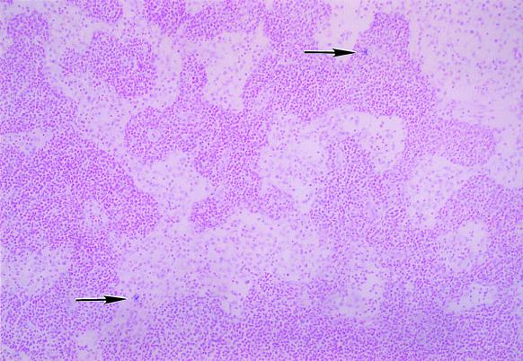
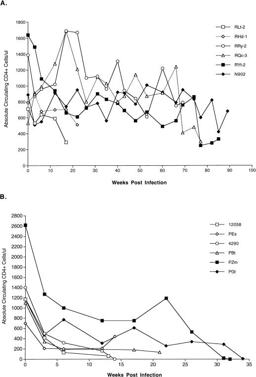
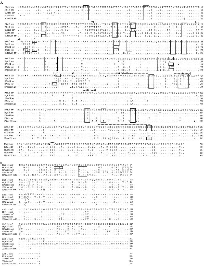
Transfusion of blood from a simian immunodeficiency virus (SIV)- and simian T-cell lymphotropic virus-infected sooty mangabey (designated FGb) to rhesus and pig-tailed macaques resulted in the development of neurologic disease in addition to AIDS. To investigate the role of SIV in neurologic disease, virus was isolated from a lymph node of a pig-tailed macaque (designated PGm) and the cerebrospinal fluid of a rhesus macaque (designated ROn2) and passaged to additional macaques. SIV-related neuropathogenic effects were observed in 100% of the pig-tailed macaques inoculated with either virus. Lesions in these animals included extensive formation of SIV RNA-positive giant cells in the brain parenchyma and meninges. Based upon morphology, the majority of infected cells in both lymphoid and brain tissue appeared to be of macrophage lineage. The virus isolates replicated very well in pig-tailed and rhesus macaque peripheral blood mononuclear cells (PBMC) with rapid kinetics. Differential replicative abilities were observed in both PBMC and macrophage populations, with viruses growing to higher titers in pig-tailed macaque cells than in rhesus macaque cells. An infectious molecular clone of virus derived from the isolate from macaque PGm (PGm5.3) was generated and was shown to have in vitro replication characteristics similar to those of the uncloned virus stock. While molecular analyses of this virus revealed its similarity to SIV isolates from sooty mangabeys, significant amino acid differences in Env and Nef were observed. This virus should provide an excellent system for investigating the mechanism of lentivirus-induced neurologic disease.


Figures

FIG. 1
FIG. 1
In situ hybridization for SIV RNA in sooty mangabey FGb. An axillary lymph node from animal FGb was used for in situ hybridization studies to identify productively infected cells. Note that only a few infected cells (that stain bluish-purple with NBT [arrows]) were found within the lymph node parenchyma, typical of cells of naturally infected, SIV-positive sooty mangabeys.
FIG. 2
FIG. 2
Longitudinal analysis of Circulating CD4+ cells in SIV-infected macaques. Blood samples from rhesus macaques (A) and pig-tailed macaques (B) infected with the FGb-derived viruses ROn2/CSF and PGm/MLN were used for the enumeration of absolute circulating CD4+ cells at the indicated time points by fluorescence-activated cell sorter analysis.
FIG. 3
FIG. 3
In situ hybridization for SIV RNA in rhesus and pig-tailed macaques infected with PGm/MLN or RON2/CSF virus isolates. Representative lymph node (A, C, E, and G) and brain (B, D, F, and H) tissues from rhesus macaques RHd1 (A and B) and RLt2 (C and D) and from pig-tailed macaques PEs (E and F) and 4290 (G and H) were examined for the presence of productive SIV infection by in situ hybridization. Moderate numbers of SIV-positive cells (indicated with the bluish-purple NBT stain) are found within the paracortices of lymph nodes from infected rhesus macaques (A and C). However, infected cells were found only in the brain of one rhesus macaque, RLt2 (D). In contrast, extremely high numbers of SIV-positive cells are found within the lymph nodes (E and G) and brains (F and H) of infected pig-tailed macaques. Note the presence of SIV-positive giant cells in the meninges of pig-tailed macaque 4290 (H, top of photograph). Arrowheads indicate cells which possess the cytomorphological characteristics of macrophages and giant cells in lymph node samples or of macrophages and microglial cells in brain samples. The arrow (Fig. 3F) indicates an SIV-infected perivascular macrophage.
FIG. 4
FIG. 4
Alignment of protein sequences of PGm5.3 and other SIV isolates. Deduced amino acid sequences of Env (A) and Nef (B) were aligned with amino acid sequences of the following isolates: SIVsmmPBj14 (PBj6.6), SIVsmH4, SIVstm (clone 37.16), and SIVmac239. For Env alignments, boxed areas indicate predicted N-linked glycosylation sites, asterisks indicate N-linked glycosylation sites in PGm5.3 not observed in other viruses, and the oval indicates a 3-amino-acid deletion in PGm5.3. For Nef alignments, boxes with solid lines indicate SH2 binding sites, boxes with dotted lines indicate conserved amino acids not present in PGm5.3, and boxes with dashed lines indicate SH3 binding sites.
FIG. 5
FIG. 5
Replication of SIVsmmFGb-derived viruses in PBMC. ConA-stimulated PBMC (107) obtained from pig-tailed macaques (A) or rhesus macaques (B) were infected with virus isolated from either ROn2/CSF, PGm/MLN, or virus derived from the molecular clone PGm5.3. Cell-free supernatants were harvested at the indicated time points and used for quantitation of RT activity as described in Materials and Methods. 32-P, 32P.

References

    1. Adamson D C, Dawson T M, Zink M C, Clements J E, Dawson V L. Neurovirulent simian immunodeficiency virus infection induces neuronal, endothelial, and glial apoptosis. Mol Med. 1996;2:417–428. - PMC - PubMed
    1. Agy M B, Frumkin L R, Corey L, Coombs R W, Wolinsky S M, Koehler J, Morton W R, Katze M G. Infection of Macaca nemestrina by human immunodeficiency virus type-1. Science. 1992;257:103–106. - PubMed
    1. Ahmed-Ansari A, Brodie A R, Fultz P N, Anderson D C, Sell K W, McClure H M. Flow microfluorometric analysis of peripheral blood mononuclear cells from nonhuman primates: correlation of phenotype with immune function. Am J Primatol. 1989;17:107–131. - PubMed
    1. Albert J, Gaines H, Sonnerborg A, Nystrom G, Pehrson P O, Chiodi F, van Sydow M, Moberg L, Lidman K, Christensson B, Asjo B, Fenyo E M. Isolation of the human immunodeficiency virus (HIV) from plasma during primary HIV infection. J Med Virol. 1987;23:67–73. - PubMed
    1. Calenda V, Chermann J C. The effects of HIV on hematopoiesis. Eur J Haematol. 1992;48:181–186. - PubMed

Publication types

MeSH terms

Associated data

LinkOut - more resources