. 1998 Nov;72(11):8893-903.
doi: 10.1128/JVI.72.11.8893-8903.1998.

Induction of programmed cell death by parvovirus H-1 in U937 cells: connection with the tumor necrosis factor alpha signalling pathway

Affiliations

Induction of programmed cell death by parvovirus H-1 in U937 cells: connection with the tumor necrosis factor alpha signalling pathway

B Rayet et al. J Virol. 1998 Nov.

Abstract

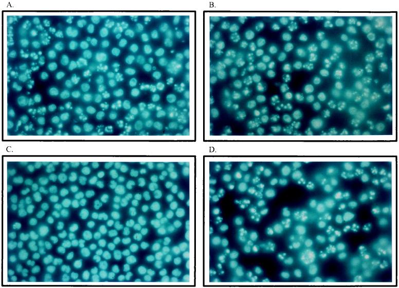
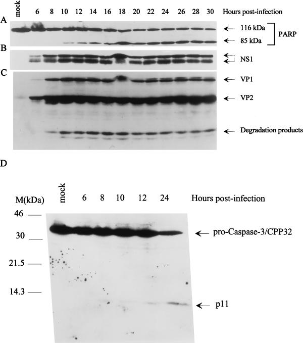
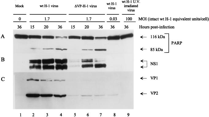
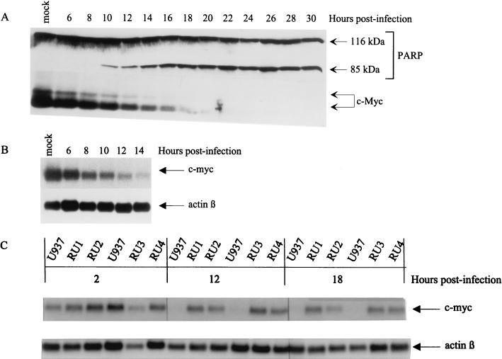
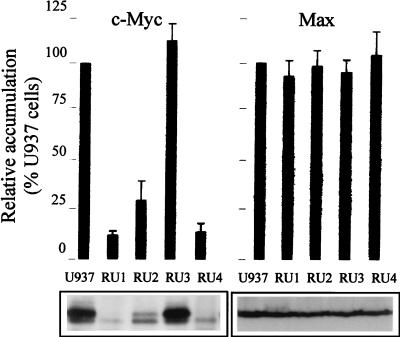
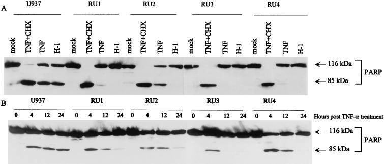
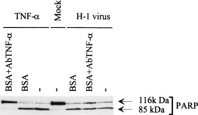
The human promonocytic cell line U937 undergoes apoptosis upon treatment with tumor necrosis factor alpha (TNF-alpha). This cell line has previously been shown to be very sensitive to the lytic effect of the autonomous parvovirus H-1. Parvovirus infection leads to the activation of the CPP32 ICE-like cysteine protease which cleaves the enzyme poly(ADP-ribose)polymerase and induces morphologic changes that are characteristic of apoptosis in a way that is similar to TNF-alpha treatment. This effect is also observed when the U937 cells are infected with a recombinant H-1 virus which expresses the nonstructural (NS) proteins but in which the capsid genes are replaced by a reporter gene, indicating that the induction of apoptosis can be assigned to the cytotoxic nonstructural proteins in this cell system. The c-Myc protein, which is overexpressed in U937 cells, is rapidly downregulated during infection, in keeping with a possible role of this product in mediating the apoptotic cell death induced by H-1 virus infection. Interestingly, four clones (designated RU) derived from the U937 cell line and selected for their resistance to H-1 virus (J. A. Lopez-Guerrero et al., Blood 89:1642-1653, 1997) failed to decrease c-Myc expression upon treatment with differentiation agents and also resisted the induction of cell death after TNF-alpha treatment. Our data suggest that the RU clones have developed defense strategies against apoptosis, either by their failure to downregulate c-Myc and/or by activating antiapoptotic factors.


Figures

FIG. 1
FIG. 1
Induction of morphologic changes in U937 cells infected with H-1 virus. Cultures (106 cells) were infected with H-1 virus (5 PFU/cell) (A and B), mock infected (C), or treated with TNF-α (10 ng/ml) (D) and further incubated for 15 h (A) and 25 h (B, C, and D). Cells were collected by low-speed centrifugation and suspended in PBS before being fixed in 4% formalin on poly-l-lysine-coated slides, stained with Hoechst solution, and examined by fluorescence microscopy.
FIG. 2
FIG. 2
Cleavage of PARP and activation of CPP32/caspase-3 in parvovirus H-1-infected cells. Total protein extracts were prepared at different times after U937 cell infection with H-1 virus (10 PFU/cell). Aliquots (70 or 150 μg) of proteins were analyzed by Western blotting with various antibodies. (A) Monoclonal PARP antibody. The bands corresponding to full-size PARP (116 kDa) or its cleavage product (85 kDa) are indicated. (B) Polyclonal anti-NS-1 serum. The phosphorylated (upper band) and nonphosphorylated (lower band) forms of NS-1 are marked. (C) Polyclonal antiserum directed against VP-1 and VP-2 capsid proteins. (D) CPP32/caspase-3 antibody. The molecular sizes are as indicated.
FIG. 3
FIG. 3
Induction of the cleavage of PARP in the presence of parvoviral NS proteins. Western blot analyses were carried out with protein extracts prepared from U937 cells at different times after infection, with intact (lanes 2 to 4, lane 8) or UV-irradiated (lane 9) wild-type H-1 virus or with a recombinant H-1 virus in which the VP genes were replaced by the jellyfish GFP (lanes 5 to 7). MOIs are given as intact wild-type virus PFU equivalents per cell. (A) PARP cleavage. (B and C) Accumulation of the H-1 virus proteins NS-1 (B) and VP-1 and VP-2 (C).
FIG. 4
FIG. 4
Downregulation of c-Myc during H-1 virus-induced apoptosis. (A) Western blot analysis of total proteins (70 μg/lane) extracted from H-1 virus-infected U937 cells (10 PFU/cell) at different times postinfection with anti-PARP (upper panel) or anti-c-Myc (lower panel) antibodies. (B) Northern blot analysis of total RNAs extracted from H-1 virus-infected U937 cells with c-myc (upper part)- or β-actin (lower part)-specific DNA probes. (C) Northern blot analysis of total RNAs extracted from H-1 virus-infected RU and U937 cells at the indicated times postinfection with c-myc (upper part)- or β-actin (lower part)-specific DNA probes.
FIG. 5
FIG. 5
Steady-state levels of c-Myc and Max proteins in parental U937 cells and H-1 virus-resistant (RU) derivatives. (Upper panels) Densitometric quantification of c-Myc and Max accumulation, expressed as percentages of the U937 level. Average values and standard-deviation bars from three experiments are shown. (Lower panels) Western blot analysis of c-Myc and Max levels in uninfected U937 and RU cells, with 50 μg of total protein extracts.
FIG. 6
FIG. 6
Comparison of the sensitivity of U937 and RU cells to TNF-α- or H-1 virus-induced apoptosis. PARP cleavage was revealed by Western blot analysis of the total protein extracts (50 μg). (Upper panel) U937 and RU cells were treated or mock treated for 24 h with TNF-α (50 ng/ml) alone, with TNF-α (50 ng/ml) plus cycloheximide (CHX) (10 μg/ml), or with H-1 virus (10 PFU/cell) as indicated at the top of each lane. (Lower panel) Cells were treated with TNF-α (1 ng/ml) for the indicated times.
FIG. 7
FIG. 7
Resistance of H-1 virus-induced apoptosis to blocking anti-TNF-α antibodies. PARP cleavage was analyzed by Western blotting with total protein extracts (50 μg) of U937 cells that were infected with H-1 virus (5 PFU/cell), mock treated, or treated with TNF-α (10 ng/ml) and further incubated for 24 h in the presence or absence of blocking anti-TNF-α serum. BSA (0.1%) was used as a negative control.

References

    1. Almasan A, Yin Y, Kelly R E, Lee E Y, Bradley A, Li W, Bertino J R, Wahl G M. Deficiency of retinoblastoma protein leads to inappropriate S-phase entry, activation of E2F-responsive genes, and apoptosis. Proc Natl Acad Sci USA. 1995;92:5436–5440. - PMC - PubMed
    1. Amati B, Land H. Myc-Max-Mad: a transcription factor network controlling cell cycle progression, differentiation and death. Curr Opin Genet Dev. 1994;4:102–108. - PubMed
    1. Askew D S, Ashmun R A, Simmons B C, Cleveland J L. Constitutive c-myc expression in an IL-3-dependent myeloid cell line suppresses cell cycle arrest and accelerates apoptosis. Oncogene. 1991;6:1915–1922. - PubMed
    1. Beg A A, Baltimore D. An essential role for NF-κB in preventing TNF-α-induced cell death. Science. 1996;274:782–784. - PubMed
    1. Beidler D R, Tewari M, Friesen P D, Poirier G, Dixit V M. The baculovirus p35 protein inhibits Fas- and tumor necrosis factor-induced apoptosis. J Biol Chem. 1995;270:16526–16528. - PubMed

Publication types