. 1998 Nov;72(11):8952-60.
doi: 10.1128/JVI.72.11.8952-8960.1998.

Tat protein induces human immunodeficiency virus type 1 (HIV-1) coreceptors and promotes infection with both macrophage-tropic and T-lymphotropic HIV-1 strains

Affiliations

Tat protein induces human immunodeficiency virus type 1 (HIV-1) coreceptors and promotes infection with both macrophage-tropic and T-lymphotropic HIV-1 strains

L Huang et al. J Virol. 1998 Nov.

Abstract

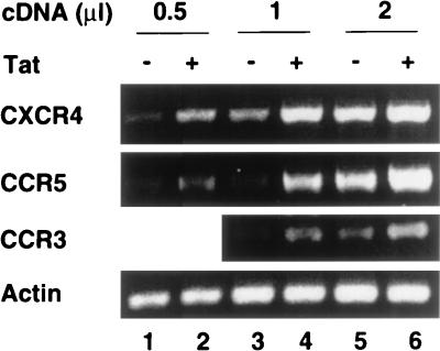
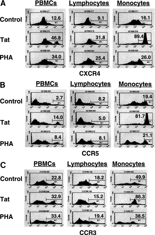
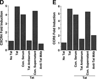
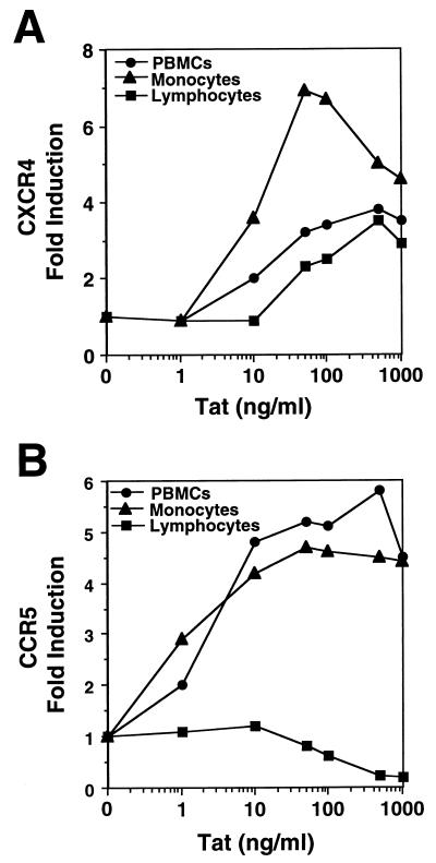
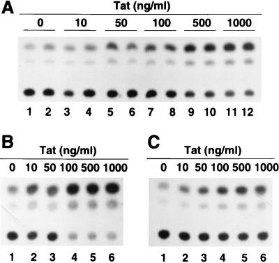
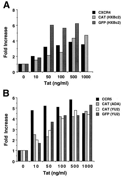
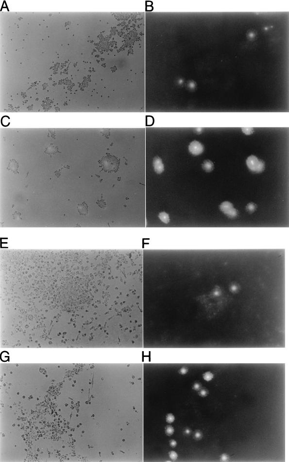
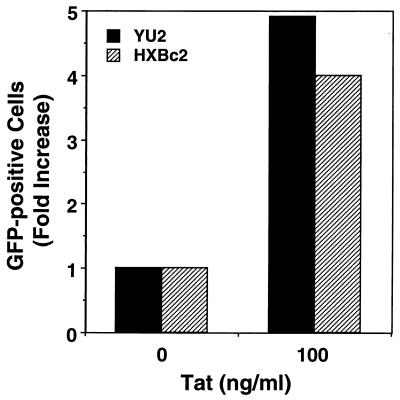
Chemokine receptors CCR5 and CXCR4 are the primary fusion coreceptors utilized for CD4-mediated entry by macrophage (M)- and T-cell line (T)-tropic human immunodeficiency virus type 1 (HIV-1) strains, respectively. Here we demonstrate that HIV-1 Tat protein, a potent viral transactivator shown to be released as a soluble protein by infected cells, differentially induced CXCR4 and CCR5 expression in peripheral blood mononuclear cells. CCR3, a less frequently used coreceptor for certain M-tropic strains, was also induced. CXCR4 was induced on both lymphocytes and monocytes/macrophages, whereas CCR5 and CCR3 were induced on monocytes/macrophages but not on lymphocytes. The pattern of chemokine receptor induction by Tat was distinct from that by phytohemagglutinin. Moreover, Tat-induced CXCR4 and CCR5 expression was dose dependent. Monocytes/macrophages were more susceptible to Tat-mediated induction of CXCR4 and CCR5 than lymphocytes, and CCR5 was more readily induced than CXCR4. The concentrations of Tat effective in inducing CXCR4 and CCR5 expression were within the picomolar range and close to the range of extracellular Tat observed in sera from HIV-1-infected individuals. The induction of CCR5 and CXCR4 expression correlated with Tat-enhanced infectivity of M- and T-tropic viruses, respectively. Taken together, our results define a novel role for Tat in HIV-1 pathogenesis that promotes the infectivity of both M- and T-tropic HIV-1 strains in primary human leukocytes, notably in monocytes/macrophages.


Figures

FIG. 1
FIG. 1
Tat induces CXCR4, CCR5, and CCR3 mRNA expression in PBMCs. Total RNA, isolated from PBMCs cultured for 3 days in the presence (+) or absence (−) of Tat (100 ng/ml), was analyzed for the expression of chemokine receptor mRNA by RT-PCR. A series of increasing amounts of cDNA, 0.5 (lanes 1 and 2), 1 (lanes 3 and 4), and 2 (lanes 5 and 6) μl from a total of 30 μl of cDNA synthesized from 2 μg of total RNA were subjected to PCR amplification. RT-PCR of β-actin mRNA was included and served as a loading control. The amplified PCR products (1,023 bp for CXCR4, 1,115 bp for CCR5, 321 bp for CCR3, and 394 bp for β-actin) are shown.
FIG. 2
FIG. 2
Tat induces CXCR4, CCR5, and CCR3 surface protein expression in PBMCs. (A through C) Indirect immunofluorescence staining and FACScan analysis of CXCR4 (A), CCR5 (B), and CCR3 (C) expression in PBMCs cultured in the absence (control) or presence of Tat (100 ng/ml) or PHA (1 μg/ml) for 4 days were carried out as described in Materials and Methods. Lymphocytes were gated according to forward and side scatter, and monocytes/macrophages were gated by using CD14 surface labeling. Percentages of positive cells were set based on negative controls (not shown) and are indicated. (D and E) Inhibition of Tat-induced up-regulation of CXCR4 (D) and CCR5 (E) expression in PBMCs by anti-Tat rabbit antiserum and anti-Tat MAb. Tat was incubated with Tat antiserum or MAb before addition to PBMC cultures. Tat pretreated with control (Con.) rabbit serum and Tat pretreated with control hybridoma supernatant were also included as controls for pretreatment with Tat antiserum or MAb. Fold induction of CXCR4 and CCR5 expression in Tat-treated PBMCs was obtained by comparison with expression in untreated cells, which was set to 1. Data are mean values of duplicates. Results shown are representative of three independent experiments.
FIG. 2
FIG. 2
Tat induces CXCR4, CCR5, and CCR3 surface protein expression in PBMCs. (A through C) Indirect immunofluorescence staining and FACScan analysis of CXCR4 (A), CCR5 (B), and CCR3 (C) expression in PBMCs cultured in the absence (control) or presence of Tat (100 ng/ml) or PHA (1 μg/ml) for 4 days were carried out as described in Materials and Methods. Lymphocytes were gated according to forward and side scatter, and monocytes/macrophages were gated by using CD14 surface labeling. Percentages of positive cells were set based on negative controls (not shown) and are indicated. (D and E) Inhibition of Tat-induced up-regulation of CXCR4 (D) and CCR5 (E) expression in PBMCs by anti-Tat rabbit antiserum and anti-Tat MAb. Tat was incubated with Tat antiserum or MAb before addition to PBMC cultures. Tat pretreated with control (Con.) rabbit serum and Tat pretreated with control hybridoma supernatant were also included as controls for pretreatment with Tat antiserum or MAb. Fold induction of CXCR4 and CCR5 expression in Tat-treated PBMCs was obtained by comparison with expression in untreated cells, which was set to 1. Data are mean values of duplicates. Results shown are representative of three independent experiments.
FIG. 3
FIG. 3
Tat-induced CXCR4 and CCR5 expression is dose dependent. CXCR4 (A) and CCR5 (B) expression was determined by indirect immunofluorescence staining of PBMCs cultured in the presence of a series of increasing concentrations of Tat for 4 days. Lymphocytes and monocytes/macrophages were gated as described in the Fig. 2 legend. The fold induction of CXCR4 and CCR5 expression in Tat-treated cells was obtained by comparison to expression in untreated cells. Data shown are representative of three independent experiments.
FIG. 4
FIG. 4
Tat promotes the infectivity of CAT reporter HIV-1 recombinant viruses. PBMCs were cultured in the presence of increasing concentrations of Tat for 4 days, infected with equal numbers of RT units of CAT reporter recombinant viruses containing HXBc2 (A), ADA (B), or YU2 (C) Env proteins, and analyzed for CAT activity at 48 h postinfection. The experiments were repeated three times, and comparable results were obtained.
FIG. 5
FIG. 5
Tat-induced CXCR4 and CCR5 expression correlates with Tat-enhanced infectivity of both CAT and GFP reporter HIV-1 recombinant viruses. CXCR4 (A) and CCR5 (B) expression was analyzed by indirect immunofluorescence staining of PBMCs cultured in the presence of a series of increasing concentrations of Tat protein for 4 days. The fold increase in the percentage of CXCR4- or CCR5-positive cells in Tat-treated PBMCs was obtained by comparison to untreated cells. The CAT assay results with CAT reporter HIV-1 viruses containing either the T-tropic (HXBc2) (A) or the M-tropic (ADA or YU2) (B) Env proteins were obtained from the autoradiograms shown in Fig. 4, which were scanned with a densitometer. The CAT activity was expressed as the percent chloramphenicol conversion. The fold increase in CAT activity in Tat-treated PBMCs was calculated by comparison to untreated cells. The GFP results were obtained by infecting PBMCs with GFP reporter HIV-1 viruses containing either HXBc2 (A) or YU2 (B) Env proteins for 9 days and analyzing the GFP fluorescence of intact cells with a flow cytometer. The fold increase in the percentage of GFP-positive cells among Tat-treated PBMCs was calculated by comparison to untreated cells.
FIG. 6
FIG. 6
Tat promoted the infectivity of the GFP reporter HIV-1 recombinant viruses in monocytes/macrophages. PBMCs were cultured in the absence (A, B, E, and F) or presence (C, D, G, and H) of Tat (100 ng/ml) for 4 days and were infected with GFP reporter HIV-1 viruses containing the M-tropic (YU2) (A through D) or T-tropic (HXBc2) (E through H) Env proteins for 9 days. After removal of the medium, the adherent cells, largely containing monocytes and monocyte-derived macrophages, were washed off nonadherent cells, fixed, and visualized by fluorescence microscopy to detect GFP fluorescence. Shown here are representative pictures depicting phase-contrast (A, C, E, and G) and fluorescence (B, D, F, and H) imaging. Results obtained from cells infected for 5 to 7 days were comparable to these.
FIG. 7
FIG. 7
The same adherent monocytes/macrophages infected with GFP reporter HIV-1 recombinant viruses shown in Fig. 6 were subsequently analyzed for GFP fluorescence by FACScan analysis. The fold increase in GFP-positive cells after treatment with Tat was obtained by comparison to untreated cells.

Similar articles

Cited by

References

    1. Alkhatib G, Combadiere C, Broder C C, Feng Y, Kennedy P E, Murphy P M, Berger E A. CC CKR5: a RANTES, MIP-1 alpha, MIP-1 beta receptor as a fusion cofactor for macrophage-tropic HIV-1. Science. 1996;272:1955–1958. - PubMed
    1. Barillari G, Gendelman R, Gallo R C, Ensoli B. The Tat protein of human immunodeficiency virus type 1, a growth factor for AIDS Kaposi sarcoma and cytokine-activated vascular cells, induces adhesion of the same cell types by using integrin receptors recognizing the RGD amino acid sequence. Proc Natl Acad Sci USA. 1993;90:7941–7945. - PMC - PubMed
    1. Berson J F, Long D, Doranz B J, Rucker J, Jirik F R, Doms R W. A seven-transmembrane domain receptor involved in fusion and entry of T-cell-tropic human immunodeficiency virus type 1 strains. J Virol. 1996;70:6288–6295. - PMC - PubMed
    1. Bhardwaj R, Becher E, Mahnke K, Hartmeyer M, Schwarz T, Scholzen T, Luger T A. Evidence for the differential expression of the functional alpha-melanocyte-stimulating hormone receptor MC-1 on human monocytes. J Immunol. 1997;158:3378–3384. - PubMed
    1. Bleul C C, Wu L, Hoxie J A, Springer T A, Mackay C R. The HIV coreceptors CXCR4 and CCR5 are differentially expressed and regulated on human T lymphocytes. Proc Natl Acad Sci USA. 1997;94:1925–1930. - PMC - PubMed

Publication types

MeSH terms