. 1998 Nov;72(11):8961-70.
doi: 10.1128/JVI.72.11.8961-8970.1998.

In vivo footprinting of the enhancer sequences in the upstream long terminal repeat of Moloney murine leukemia virus: differential binding of nuclear factors in different cell types

Affiliations

In vivo footprinting of the enhancer sequences in the upstream long terminal repeat of Moloney murine leukemia virus: differential binding of nuclear factors in different cell types

S W Granger et al. J Virol. 1998 Nov.

Abstract

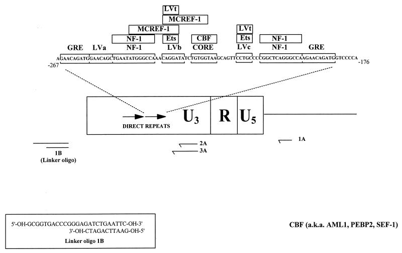
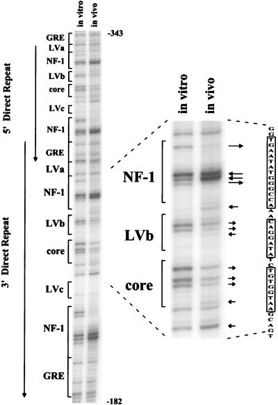
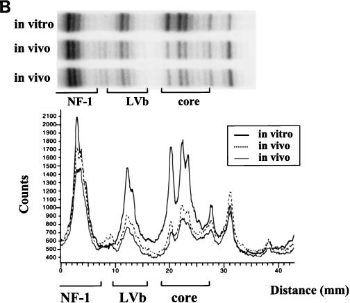
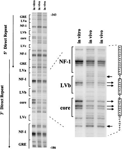
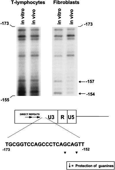
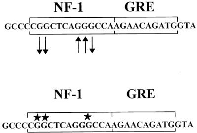
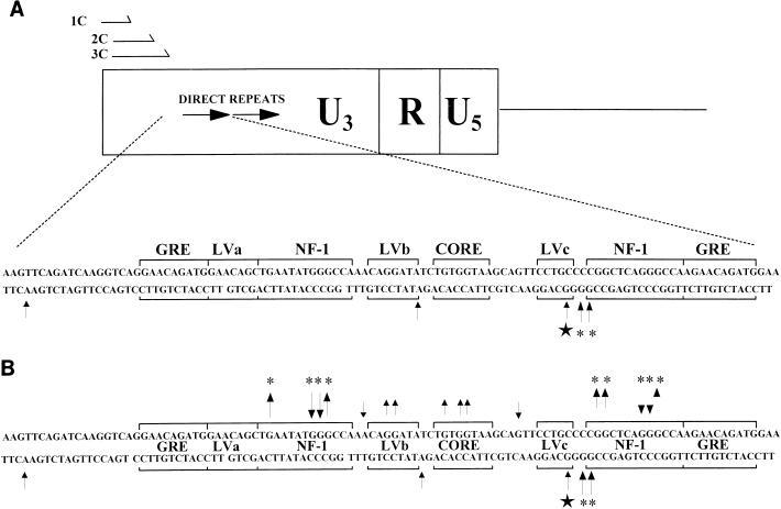
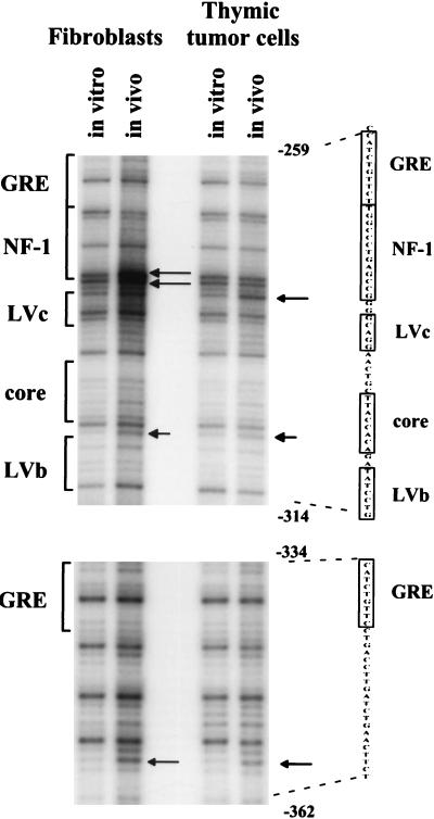
The enhancer sequences in the Moloney murine leukemia virus (M-MuLV) long terminal repeat (LTR) are of considerable interest since they are crucial for virus replication and the ability of the virus to induce T lymphomas. While extensive studies have identified numerous nuclear factors that can potentially bind to M-MuLV enhancer DNA in vitro, it has not been made clear which of these factors are bound in vivo. To address this problem, we carried out in vivo footprinting of the M-MuLV enhancer in infected cells by in vivo treatment with dimethyl sulfate (DMS) followed by visualization through ligation-mediated PCR (LMPCR) and gel electrophoresis. In vivo DMS-LMPCR footprinting of the upstream LTR revealed evidence for factor binding at several previously characterized motifs. In particular, protection of guanines in the central LVb/Ets and Core sites within the 75-bp repeats was detected in infected NIH 3T3 fibroblasts, Ti-6 lymphoid cells, and thymic tumor cells. In contrast, factor binding at the NF-1 sites was found in infected fibroblasts but not in T-lymphoid cells. These results are consistent with the results of previous experiments indicating the importance of the LVb/Ets and Core sequences for many retroviruses and the biological importance especially of the NF-1 sites in fibroblasts and T-lymphoid cells. No evidence for factor binding to the glucocorticoid responsive element and LVa sites was found. Additional sites of protein binding included a region in the GC-rich sequences downstream of the 75-bp repeats (only in fibroblasts), a hypersensitive guanine on the minus strand in the LVc site (only in T-lymphoid cells), and a region upstream of the 75-bp repeats. These experiments provide concrete evidence for the differential in vivo binding of nuclear factors to the M-MuLV enhancers in different cell types.


Figures

FIG. 1
FIG. 1
Nuclear factor binding sites in the M-MuLV enhancers. A schematic representation of the M-MuLV LTR with the nuclear protein binding sites (bracketed sequences) and sequence-specific DNA proteins known to bind to them (in boxes) in one copy of the 75-bp direct repeats is shown. (These data are taken from Manley et al. [26]). The nested oligonucleotide primer set (1A, 2A, and 3A) used for LMPCR is shown below. Oligonucleotide 1A enabled preferential analysis of the 5′ LTR without interference from signal due to binding sites at the 3′ LTR. The sequence of the linker oligonucleotide used in the LMPCR (1B) is also shown. Oligo, oligonucleotide.
FIG. 2
FIG. 2
In vivo DMS footprinting of the M-MuLV 5′ LTR in fibroblasts. An autoradiogram from gel electrophoresis of labeled LMPCR products obtained from M-MuLV-infected 43-D fibroblasts is shown. The autoradiogram is representative of multiple analyses with the same nested oligonucleotide primer set. The portion of the gel shown corresponds to nucleotides −182 to −343 in the upstream M-MuLV LTR. The relative positions of previously characterized nuclear protein binding sites are indicated on the left. Both 75-bp direct repeats of the M-MuLV enhancer are displayed, and the region of interest is expanded at the right. The boxed sequences at the far right correspond to the NF-1, LVb, and Core sites. Comparisons between the in vitro-DMS-treated DNA control in the lane to the left and the in vivo-DMS-treated sample in the lane to the right indicated protection of certain guanine bases and hypersensitivity of other guanine bases in the infected cells. Guanine-specific protection is indicated by arrows pointing away from bands, and guanine or adenine hypersensitivity to DMS is indicated by arrows pointing towards the bands. Other investigators have previously reported hypersensitivity of adenines in in vivo DMS-LMPCR footprinting. The lengths of the arrows indicate the relative magnitudes of the protection or hypersensitivity.
FIG. 3
FIG. 3
In vivo DMS footprinting of the M-MuLV 5′ LTR in infected lymphoid cells. (A) In vivo DMS-LMPCR footprinting analogous to that described for Fig. 2 was carried out with M-MuLV-infected Ti-6 lymphoid cells. Infected Ti-6 cells were subjected to LMPCR after two independent in vivo DMS treatments. In this figure, the LMPCR fragments were visualized by PhosphorImaging. The same convention as that described for Fig. 2 was used, with arrows indicating protected and hypersensitive sites. (B) Digital densitometric analysis of the PhosphorImaged sequencing gel in panel A was performed. The positions of the NF-1, LVb/Ets, and Core sites are indicated. The portion of the Ti-6 gel analyzed is aligned above the graph.
FIG. 3
FIG. 3
In vivo DMS footprinting of the M-MuLV 5′ LTR in infected lymphoid cells. (A) In vivo DMS-LMPCR footprinting analogous to that described for Fig. 2 was carried out with M-MuLV-infected Ti-6 lymphoid cells. Infected Ti-6 cells were subjected to LMPCR after two independent in vivo DMS treatments. In this figure, the LMPCR fragments were visualized by PhosphorImaging. The same convention as that described for Fig. 2 was used, with arrows indicating protected and hypersensitive sites. (B) Digital densitometric analysis of the PhosphorImaged sequencing gel in panel A was performed. The positions of the NF-1, LVb/Ets, and Core sites are indicated. The portion of the Ti-6 gel analyzed is aligned above the graph.
FIG. 4
FIG. 4
In vivo DMS footprinting of the M-MuLV 5′ LTR in primary M-MuLV-induced tumor cells. M-MuLV-induced T-lymphoma cells were treated in vivo with DMS and analyzed in the same manner as described for the 43-D and infected Ti-6 cells shown in Fig. 2 and 3. PhosphorImaging was employed to visualize data. In this experiment, the intensity of the entire in vitro lane was uniformly decreased to equalize the amounts of samples loaded and to aid in footprint visualization.
FIG. 5
FIG. 5
In vivo DMS footprinting in the GC-rich sequences. The regions from DMS footprinting gels analogous to those shown in Fig. 2 and 4 and corresponding to the GC-rich sequences downstream of the 75-bp repeats are shown for thymic tumor DNA and 43-D infected fibroblasts. Evidence for protein interactions at positions −153 and −155 in the sequence CAGCAG was obtained for fibroblasts but not for M-MuLV-infected Ti-6 cells or the primary thymic tumor cells. Infected Ti-6 cells showed the same pattern as the thymic tumor cells (no protection [data not shown]). The relative positions of the protected bases in the GC-rich sequences are shown.
FIG. 6
FIG. 6
Summary of DNA-protein interactions on the sense strand obtained by in vivo footprinting by LMPCR. The same convention described for Fig. 2 was used to summarize all interactions observed in Fig. 2 to 5. Arrows in the LVb-Core region represent interactions observed in both lymphoid cells and fibroblasts infected with M-MuLV; those pointing upward indicate sites hyperactive to DMS, and those pointing downward indicate protection of specific guanines. Other interactions were observed only in M-MuLV-infected fibroblasts.
FIG. 7
FIG. 7
Comparison of in vivo footprints and in vitro methylation interference results. Nuclear protein interactions at the NF-1 site reported by Speck and Baltimore (40) detected by methylation interference with WEHI 231 nuclear extracts are shown in the lower sequence (stars indicate G residues that interacted with protein). The results of in vivo DMS footprinting are shown in the upper sequence. Guanine bases protected in vivo (arrows pointing away from bases) correspond to guanines shown to be protein contact points by methylation interference.
FIG. 8
FIG. 8
DNA-protein interactions on the minus strand obtained by in vivo footprinting. (A) The nested oligonucleotide primer set used for the minus-strand in vivo footprinting (1C, 2C, and 3C) is shown. A summary of the minus (lower)-strand bases hypersensitive to in vivo DMS methylation in fibroblasts and primary thymic tumor cells was compiled analogously to that shown in Fig. 6. These results were taken from the in vivo footprinting analysis shown in Fig. 9. Asterisks indicate bases hypersensitive to DMS methylation in infected fibroblasts, the star indicates a hypersensitive base specific to primary thymic tumor cells, and arrows indicate sites hyperactive to DMS. (B) A summary of in vivo protein-DNA interactions observed for both DNA strands is shown. Arrows pointing away from bases on either strand indicate DMS protection, while arrows pointing towards bases indicate DMS-hypersensitive sites. Note that protein interactions on the minus strand were evident only as hypersensitive sites. The data for the sense (upper) strand are specific for the upstream LTR, while the data for the minus strand are composites of the upstream and downstream LTRs. Stars indicate T-lymphoid-cell-specific bases, and asterisks indicate fibroblast-specific bases.
FIG. 9
FIG. 9
In vivo footprinting of the M-MuLV LTR minus strand. Infected 43-D fibroblasts and thymic tumor cells treated in vivo or in vitro with DMS were analyzed by cleavage and LMPCR by using the minus-strand-specific primers shown in Fig. 8A. Arrows indicate bases hypersensitive to in vivo DMS methylation in each respective cell type. DNA templates were the same as those used for the sense-strand analysis.

Similar articles

Cited by

References

    1. Boral A L, Okenquist S A, Lenz J. Identification of the SL3-3 virus enhancer core as a T-lymphoma cell-specific element. J Virol. 1989;63:76–84. - PMC - PubMed
    1. Chatis P A, Holland C A, Hartley J W, Rowe W P, Hopkins N. Role for the 3′ end of the genome in determining disease specificity of Friend and Moloney murine leukemia viruses. Proc Natl Acad Sci USA. 1983;80:4408–4411. - PMC - PubMed
    1. Chatis P A, Holland C A, Silver J E, Frederickson T N, Hopkins N, Hartley J W. A 3′ end fragment encompassing the transcriptional enhancers of nondefective Friend virus confers erythroleukemogenicity on Moloney leukemia virus. J Virol. 1984;52:248–254. - PMC - PubMed
    1. Chen H, Yoshimura F K. Identification of a region of a murine leukemia virus long terminal repeat with novel transcriptional regulatory activities. J Virol. 1994;68:3308–3316. - PMC - PubMed
    1. DesGroseillers L, Jolicoeur P. Mapping the viral sequences conferring leukemogenicity and disease specificity in Moloney and amphotropic murine leukemia viruses. J Virol. 1984;52:448–456. - PMC - PubMed

Publication types

MeSH terms