. 1998 Nov;72(11):8988-9001.
doi: 10.1128/JVI.72.11.8988-9001.1998.

African swine fever virus is enveloped by a two-membraned collapsed cisterna derived from the endoplasmic reticulum

Affiliations

African swine fever virus is enveloped by a two-membraned collapsed cisterna derived from the endoplasmic reticulum

G Andrés et al. J Virol. 1998 Nov.

Abstract

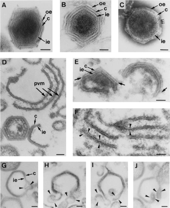
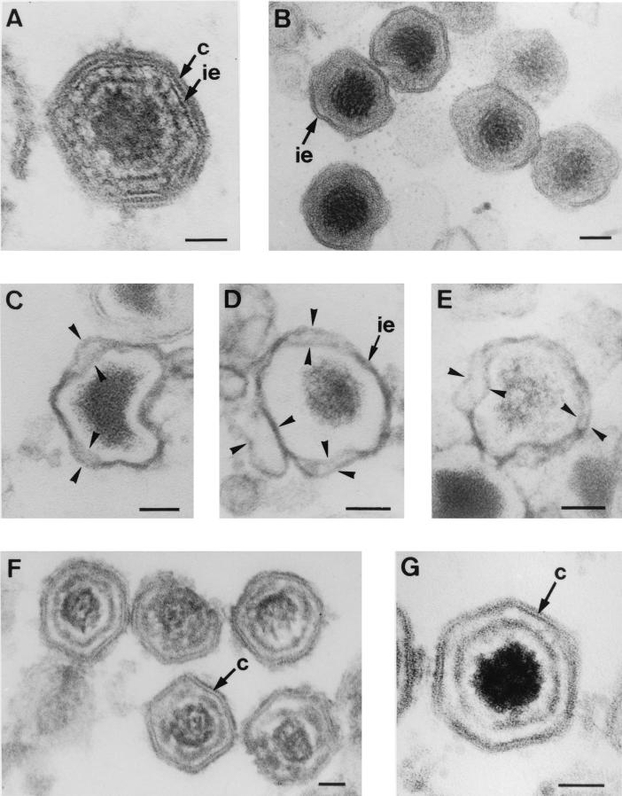
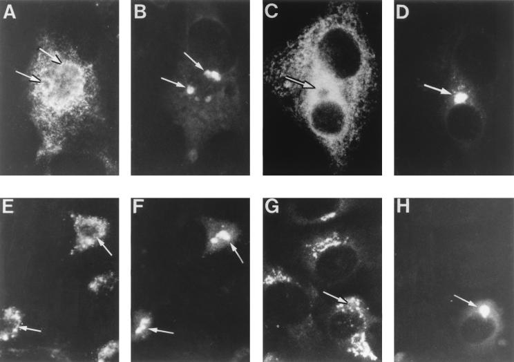
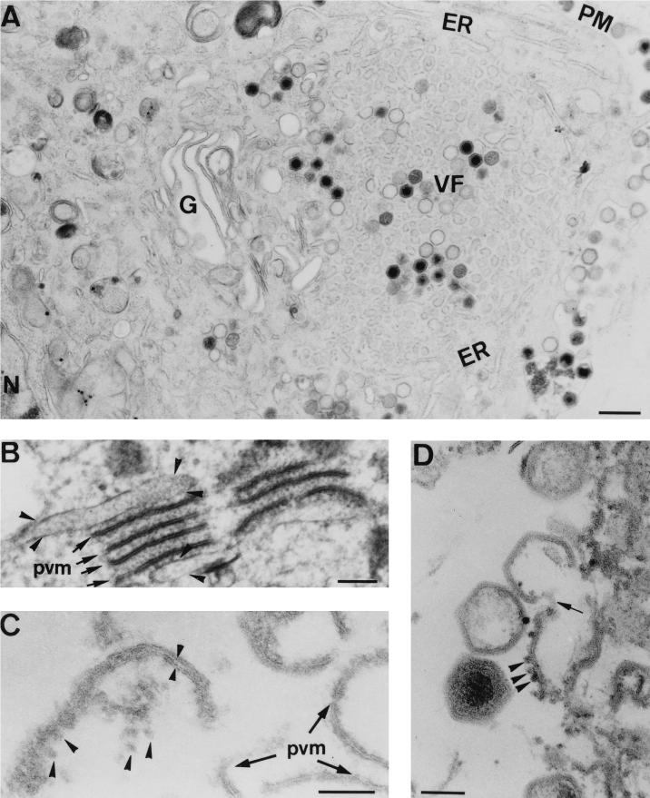
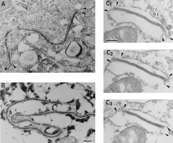
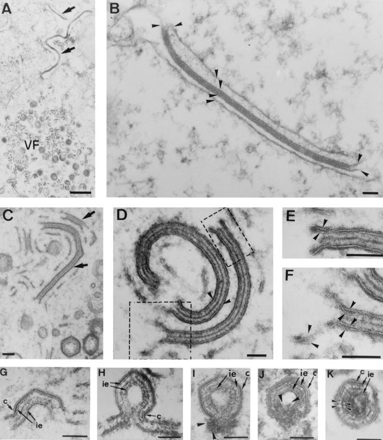
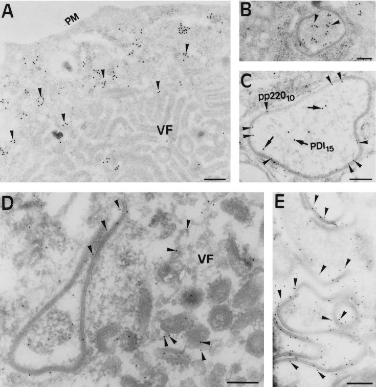
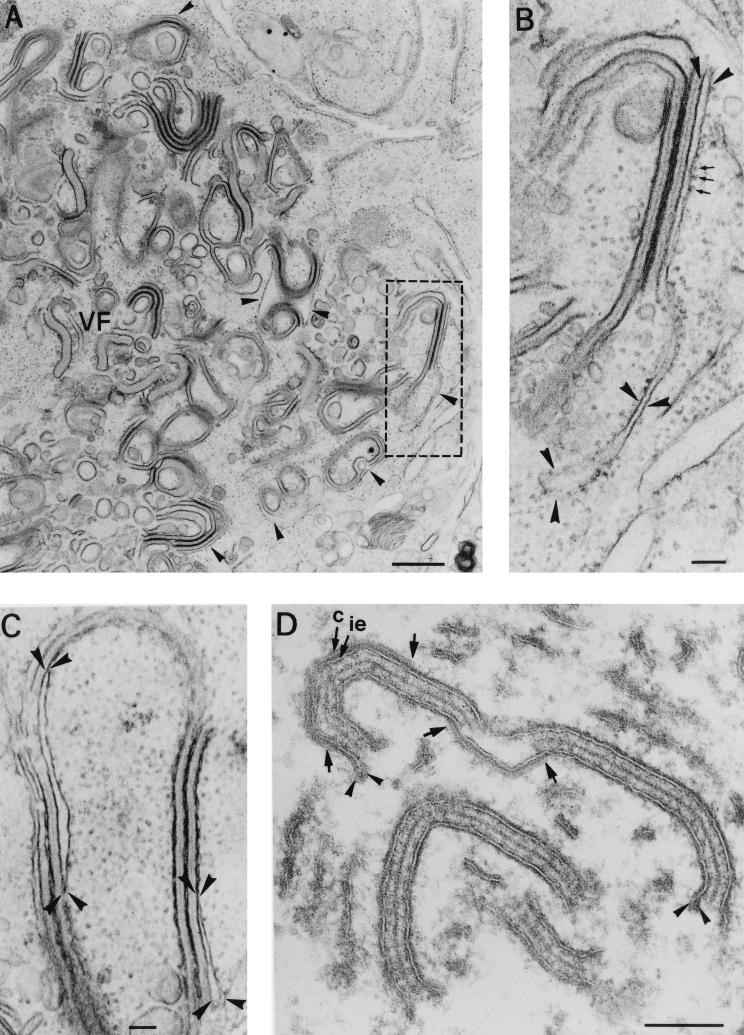
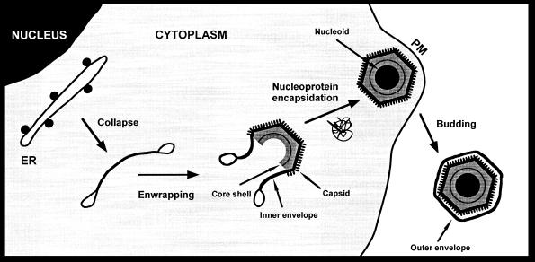
During the cytoplasmic maturation of African swine fever virus (ASFV) within the viral factories, the DNA-containing core becomes wrapped by two shells, an inner lipid envelope and an outer icosahedral capsid. We have previously shown that the inner envelope is derived from precursor membrane-like structures on which the capsid layer is progressively assembled. In the present work, we analyzed the origin of these viral membranes and the mechanism of envelopment of ASFV. Electron microscopy studies on permeabilized infected cells revealed the presence of two tightly apposed membranes within the precursor membranous structures as well as polyhedral assembling particles. Both membranes could be detached after digestion of intracellular virions with proteinase K. Importantly, membrane loop structures were observed at the ends of open intermediates, which suggests that the inner envelope is derived from a membrane cisterna. Ultraestructural and immunocytochemical analyses showed a close association and even direct continuities between the endoplasmic reticulum (ER) and assembling virus particles at the bordering areas of the viral factories. Such interactions become evident with an ASFV recombinant that inducibly expresses the major capsid protein p72. In the absence of the inducer, viral morphogenesis was arrested at a stage at which partially and fully collapsed ER cisternae enwrapped the core material. Together, these results indicate that ASFV, like the poxviruses, becomes engulfed by a two-membraned collapsed cisterna derived from the ER.


Figures

FIG. 1
FIG. 1
Structure and assembly of ASFV. (A to C) Extracellular ASFV particles processed by Epon sectioning (A), cryosectioning (B), and negative staining (C). All these EM methods reveal an overall structure consisting of a central core surrounded by three layers: the inner envelope (ie), the capsid (c), and the outer envelope (oe). Note that in the negatively stained particle, individual capsomers (small arrows in panel C) are evident within the capsid. (D to J) Ultrathin Epon sections of viral intermediates from infected Vero cells permeabilized at 18 to 24 h p.i. Intracellular ASFV particles mature from precursor viral membranous structures (pvm) present in the viral factories (D), which become polyhedral particles by the gradual formation of the capsid on one of their faces (D and E). The short arrows in panel E indicate the limits of capsids assembling on the inner envelopes. Note also the electron-dense core material underneath the concave side of the inner envelope (E). Close inspection of the assembly intermediates revealed two distinct membranes within the precursor membranous structures (arrowheads in panel F) as well as in polyhedral assembling particles (arrowheads in panels G to J). Note that the ends of open particles (H to J) appeared as membrane loops, which suggests that ASFV becomes enwrapped by a collapsed two-membraned cisterna. Bars, 50 nm.
FIG. 2
FIG. 2
Selective disruption of ASFV particles. (A) Epon section of an intact intracellular particle showing the central core successively enclosed by the inner envelope (ie) and the outer capsid (c). (B to E) Epon sections of intracellular virions incubated with proteinase K. At 20 h p.i. infected Vero cells were perforated by hypotonic lysis and subsequently incubated with proteinase K for 30 min at room temperature. After the proteinase treatment, intracellular particles had lost the capsid but not the inner envelope (B). Importantly, in highly damaged virions the inner envelope appeared dissociated in two distinct membranes (arrowheads in panels C to E). Note also the dramatic alteration of their icosahedral morphology. (F and G) Epon sections of purified extracellular particles treated for 30 min with the nonionic detergent β-d-octylglucopyranoside. Note the lack of the outer and the inner envelopes and the partial disruption of the viral core. Note also that the capsid remains apparently intact and the resulting particles retain their polyhedral shapes. Bars, 50 nm.
FIG. 3
FIG. 3
Immunofluorescence analysis of ER and Golgi marker proteins in ASFV-infected cells. Infected Vero cells were fixed at 12 to 16 h p.i. with methanol at −20°C. Double-labeling experiments were performed with antibodies to ASFV polyprotein pp220 to stain the viral factories (B, D, F, and H) and with antibodies to different ER and Golgi proteins (A, C, E, and G), as follows. For ER labeling, a rabbit antiserum to the luminal marker PDI (A) and a rabbit antiserum to membrane ER glycoproteins (C) were used. For Golgi labeling, a MAb to the membrane glycoprotein gp74 (E) and a rabbit serum to galactosyltransferase (G) were used. The antibodies to pp220 were a mouse MAb (24A.G4) (B, D, and H) and the rabbit antiserum anti-pp220/p150 (F). Double labeling was developed with fluorescein-coupled secondary antibodies for the assembly sites and Texas red-conjugated antibodies for the organelle markers. Note that viral factories (arrows) essentially exclude the ER and Golgi markers but are closely encompassed by them.
FIG. 4
FIG. 4
Relationship between viral membranes and ER membranes. Ultrathin Epon sections of ASFV-infected Vero cells at 24 h p.i. are shown. (A) Viral factories (VF) were usually encompassed by an enlarged Golgi complex (G) and ER cisternae (ER). The plasma membrane (PM) and the nucleus (N) are also indicated. (B to D) At the limits of the assembly sites, a close association between ER membranes and viral structures was often evident. (B) Several precursor viral membranes (pvm, arrows) are present between two cellular cisternae (arrowheads). Note the presence of electron-dense viroplasmic material associated with both viral and cellular membranes. (C) An apparently collapsed ER cisterna (small arrowheads) with attached ribosomes (large arrowheads) is in close vicinity to precursor viral membranes. (D) Eventually, direct continuities (arrow) between membranes with attached ribosomes (arrowheads) and assembling virions were also evident. Bars: 500 nm (A), 100 nm (B to D).
FIG. 5
FIG. 5
Topology of the zipper-like structures. (A and B) Ultrathin Epon sections of marginal zipper-like structures within a nonpermeabilized (A) or SLO-permeabilized (B) infected cell at 16 h p.i. Note in the control (A) how the limits of the viral intermediate appear to be continuous with two adjacent ER cellular cisternae (arrowheads). After cell permeation (B), the cytosol (C) is extracted but not the luminal contents (L), which become obvious. Thus, it is clear that the core shell is a cytosolic structure limited by two ER cisternae. (C1 to C3) Set of serial sections of a peripheral zipper-like structure. The core shell is interpreted as a laminar structure whose limiting surfaces interact with the cytoplasmic sides of ER cisternae. Note the presence of ribosomes (small arrows) attached to ER membranes (arrowheads). Bars: 200 nm (A and B) and 100 nm (C1).
FIG. 6
FIG. 6
Assembly of zipper-like structures. Epon sections of viral zipper-like structures present in the bordering areas of the assembly sites (A and B) or within them (C to I). (A and B) Outside the viral factories (VF), these atypical viral intermediates (arrows in panel A) consist of an extended core shell limited by two membrane cisternae (arrowheads in panel B). (C to E) Within the viral factories, closely related zipper-like structures are also found (arrows in panel C). These intermediates are composed by an extended core shell limited by two typical membrane-like structures (arrowheads in panel D). When examined in detail (panels E and F are higher magnifications of the areas delimited in panel D), two apposed lipid membranes (arrowheads) can be discerned within each limiting viral envelope. (G to K) Eventually, the zipper-like structures become polyhedral particles by the progressive formation of a capsid layer (c) on one of the two limiting envelopes (ie). Concomitantly, a nucleoprotein-like material appears to be encapsidated within some double-enveloped particles (arrowheads in panels I and J). Finally, after membrane fusion events, the resulting closed particles contain two inner envelopes encompassing the core shell (K). Note that the core shell is formed by two regular arrays of protein subunits (arrowheads in panel K) separated by a thin electron-dense protein layer. Bars: 500 nm (A) and 100 nm (B to K).
FIG. 7
FIG. 7
Immunogold labeling of ER marker proteins in ASFV-infected cells. Vero cells infected with ASFV for 18 h p.i. were processed by freeze-substitution (A to C) or cryosectioning (D and E). (A and B) Ultrathin sections were incubated with a MAb against the luminal ER protein PDI followed by rabbit anti-mouse immunoglobulin G and protein A-gold (diameter, 15 nm). Note that anti-PDI labeling (arrowheads) is essentially excluded from the assembly sites (A) but not from the luminal contents of peripheral zipper-like structures (B). (C) Double labeling of a zipper-like structure with antiserum to polyprotein pp220 followed by protein A-gold (diameter, 10 nm) and with the anti-PDI MAb followed by protein A-gold (diameter, 15 nm). The anti-pp220 labeling (arrowheads) is located within the core shell, while PDI (arrows) is present within the associated cisterna. (D and E) Thawed cryosections were incubated with anti-MERG antiserum followed by protein A-gold (diameter, 10 nm). The labeling is associated with the membranes of marginal zipper-like structures (D and E) and, to a much lesser extent, to viral structures within the viral factories (E). Bars: 500 nm (A) and 200 nm (B to E).
FIG. 8
FIG. 8
Origin of the inner envelope of ASFV recombinant vA72. Epon sections of Vero cells infected with recombinant virus vA72 for 18 h in the absence of IPTG (A to C), or treated with the inducer at 18 h p.i. for an 8-h period (D) are shown. (A) Under nonpermissive conditions, the viral factories (VF) show a great accumulation of zipper-like structures and a virtual absence of polyhedral viral structures, as a consequence of the inhibition of capsid formation. In the peripheral areas of the assembly sites, the zipper-like structures appear clearly associated with ER cisternae (arrowheads). (B) Higher magnification of the region delimited in panel A. Note how a rough ER cisterna (arrowheads) appears directly bound to an extended viral core shell. The small arrows indicate ribosomes. (C) Partially collapsed ER cisternae associated with zipper-like structures. The arrowheads delimit local extensions where the cisternal structure is still evident. In our interpretation, the collapse would lead to formation of the viral envelopes by the tight apposition of the two limiting lipid bilayers. (D) Detail of a viral factory after an 8-h period of IPTG induction. Under these conditions, the zipper-like structures become polyhedral intermediates by the de novo and gradual assembly of the capsid layer (c) on the inner envelope (ie). The arrows indicate the ends of two capsids assembling on opposite faces of the same zipper-like structure. Note also the presence of membrane loops (arrowheads) at the ends of the viral envelopes. Bars: 500 nm (A) and 100 nm (B to D).
FIG. 9
FIG. 9
Model for ASFV assembly. Intracellular ASFV particles acquire their inner envelopes from the ER. The envelopment probably begins by the insertion of viral proteins into the ER membranes and, concomitantly, the exclusion of the host membrane proteins. During this process, the cell compartment would be collapsed to give rise to precursor viral structures formed by two tightly apposed membranes. Subsequently, the capsid would be gradually assembled on one side of the inner envelope whereas the core shell would form beneath the opposite face. At the time the particle is closing, the nucleoprotein material of the nucleoid would become engulfed. Finally, the intracellular particles would release from the cell by budding at the plasma membrane (PM). According to this model, the resulting extracellular ASFV particles would contain three lipid membranes.

References

    1. Alcalde J, Egea G, Sandoval I V. gp74, a membrane glycoprotein of a cis-Golgi network that cycles through the endoplasmic reticulum and intermediate compartment. J Cell Biol. 1994;124:649–665. - PMC - PubMed
    1. Almeida J D, Waterson A P, Plowright W. The morphological characteristics of African swine fever virus and its resemblance to Tipula iridescent virus. Arch Gesamte Virusforsch. 1967;20:392–396. - PubMed
    1. Andrés G, Simón-Mateo C, Viñuela E. Assembly of African swine fever virus: role of polyprotein pp220. J Virol. 1997;71:2331–2341. - PMC - PubMed
    1. Breese S S, Jr, DeBoer C J. Electron microscope observation of African swine fever virus in tissue culture cells. Virology. 1966;28:420–428. - PubMed
    1. Brookes S M, Dixon L K, Parkhouse R M E. Assembly of African swine fever virus: quantitative ultrastructural analysis in vitro and in vivo. Virology. 1996;224:84–92. - PubMed

Publication types

Substances

LinkOut - more resources