Comparative Study
. 1998 Nov;72(11):9045-53.
doi: 10.1128/JVI.72.11.9045-9053.1998.

Neuronal death induced by brain-derived human immunodeficiency virus type 1 envelope genes differs between demented and nondemented AIDS patients

Affiliations
Comparative Study

Neuronal death induced by brain-derived human immunodeficiency virus type 1 envelope genes differs between demented and nondemented AIDS patients

C Power et al. J Virol. 1998 Nov.

Abstract

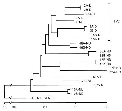
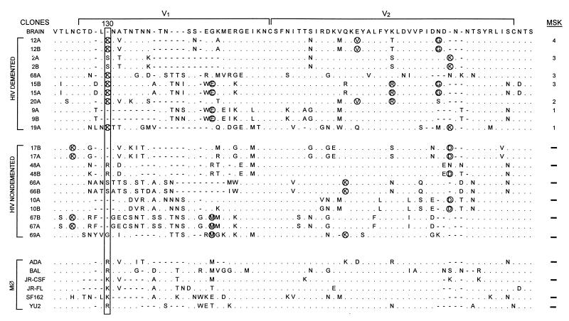
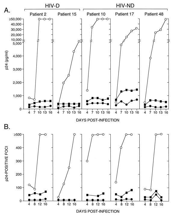
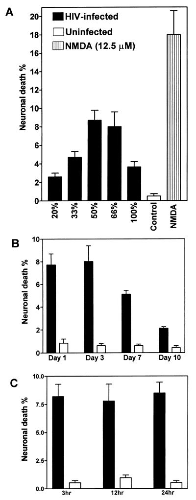
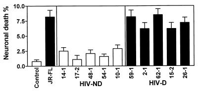
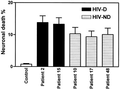
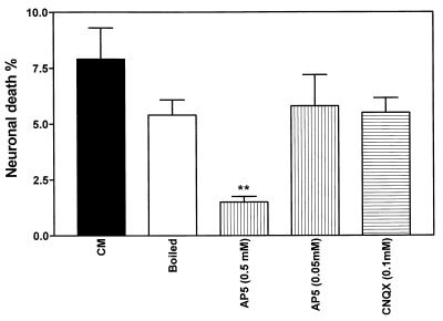
Human immunodeficiency virus type 1 (HIV-1) infection of the brain results in viral replication primarily in macrophages and microglia. Despite frequent detection of viral genome and proteins in the brains of AIDS patients with and without HIV dementia, only 20% of AIDS patients become demented. To investigate the role of viral envelope gene variation in the occurrence of dementia, we examined regions of variability in the viral envelope gene isolated from brains of AIDS patients. Brain-derived HIV-1 V1-V2 envelope sequences from seven demented and six nondemented AIDS patients displayed significant sequence differences between clinical groups, and by phylogenetic analysis, sequences from the demented group showed clustering. Infectious recombinant viruses containing brain-derived V3 sequences from both clinical groups were macrophagetropic, and viruses containing brain-derived V1, V2, and V3 sequences from both clinical groups spread efficiently in macrophages. In an indirect in vitro neurotoxicity assay using supernatant fluid from HIV-1-infected macrophages, recombinant viruses from demented patients induced greater neuronal death than viruses from nondemented patients. Thus, the HIV-1 envelope diversity observed in these patient groups appeared to influence the release of neurotoxic molecules from macrophages and might account in part for the variability in occurrence of dementia in AIDS patients.


Figures

FIG. 1
FIG. 1
The entire HIV-1 gp120 sequence (511 amino acids) showing the regions of interest, including the V1-V2 and C2-V3 fragments and restriction sites used in this study. The V1 and V2 regions lie between the DraIII and StuI sites (76 amino acids), and the C2 and V3 regions are within the StuI-to-NheI fragment (143 amino acids).
FIG. 2
FIG. 2
Phylogenetic comparison of brain-derived V1-V2 sequences obtained from HIV-D (D) and HIV-ND (ND) individuals with AIDS and the consensus sequence from clade D (con.D clade) (38), using neighbor-joining analysis (30). Numbers refer to individual patients, and letters (A and B) refer to clones from the same patient. Two clones were analyzed for each patient. Clones from five of seven HIV-D patients clustered together, while none of the HIV-ND patients showed close associations. The horizontal axis represents the number of substitution events. Analysis of the V1 and V2 sequences separately revealed no clustering of sequences from the same clinical group. Similar topologies were obtained with different phylogenetic methods, but bootstrap values were low (<70) between individuals.
FIG. 3
FIG. 3
Brain-derived V1-V2 envelope sequences from HIV-D (D) and HIV-ND (ND) patients aligned with the brain consensus sequence and corresponding sequences from established macrophagetropic viruses. HIV-D sequences are presented in order of severity of dementia according to MSK score shown at the right. Two clones were sequenced from each patient except for patients 68, 20, 19, and 69. Position 130 (boxed) shows significant difference between HIV-D and HIV-ND groups (Fisher’s exact, P < 0.01), with a lysine predominating in the HIV-D group and a variable amino acid in the HIV-ND group. Comparison of the frequency of the Lys130 in established databases (38) revealed that it was detected significantly more frequently in the HIV-D sequences than in the database (Fisher’s exact, P < 0.01). Circled amino acids identified in two or more patients in one group, but not present in the other group at a specific position, were termed unique. The mean number of unique amino acids per clone ± SEM was significantly greater in the HIV-D group (3.0 ± 0.44) than in the HIV-ND group (1.6 ± 0.20) (Student’s t test, P < 0.05). In patients with two or more clones, the difference between clones varied from zero to three amino acids. The length of the V2 region, the number of positively charged amino acids, and number of potential glycosylation sites did not differ between HIV-D and HIV-ND groups.
FIG. 4
FIG. 4
Infectivity of brain-derived C2-V3- and V1-V3-containing recombinant viruses measured by p24 levels in macrophage culture supernatant (A) or p24-positive foci (B) over time. At an input titer of 102 TCID50/0.1 ml, V1-V3 clones (open circles) replicated to a greater extent in macrophages, reflected by higher p24 levels in supernatant (A) and progressively increasing numbers of p24-positive cells (B), compared to the matched C2-V3 clones (closed circles) for both HIV-D- and HIV-ND-derived viruses. When a 10-fold-higher input of C2-V3 viruses was used (closed squares), higher p24 values and focus counts were observed for all viruses, confirming that these C2-V3 clones were macrophagetropic. The increasing p24 level in patient 17 did not reflect an increase in focus number and probably was due to accumulation of p24 in the medium, since all medium was not removed when cells were fed every third day.
FIG. 5
FIG. 5
Comparison of mean percentage (±SEM) of neuronal death induced by CM from macrophages infected with HIV-1 JR-FL, uninfected macrophages (control), or NMDA treatment (A), CM harvested from different time points after infection (B), and different times of exposure of neuronal cultures to CM (C). JR-FL induced neurotoxicity at various dilutions of CM (A), with maximum neuronal death at a 50 to 66% dilution. Neuronal cultures treated with 50% CM from uninfected macrophages showed significantly less neuronal death than CM at the same concentration from JR-FL-infected macrophages (Student’s t test, P < 0.001). A decline in neuronal killing was observed with 50% CM harvested at later time points postinfection (B). The extent of neuronal death did not change with different times of exposure to CM, harvested at 3 days postinfection (C). Macrophages were infected at an input titer of 103 TCID50/0.1 ml.
FIG. 6
FIG. 6
Mean percentage of neuronal death (±SEM) caused by CM from control (uninfected) or JR-FL-infected macrophage cultures or macrophage cultures infected with recombinant viruses containing brain-derived C2-V3 sequences from HIV-D and HIV-ND individuals. Mean neuronal death caused by all recombinant HIV-D viruses was significantly greater than neuronal death caused by all HIV-ND viruses (Student’s t test, P < 0.0001). Each HIV-D-derived virus caused significantly more neuronal death than each HIV-ND-derived virus when the isolates were compared individually (P < 0.05). Macrophages were infected at an input titer of 103 TCID50/0.1 ml.
FIG. 7
FIG. 7
Comparison of mean percentages (±SEM) of neuronal death induced by CM for brain-derived V1-V3-containing recombinant viruses from two HIV-D and three HIV-ND patients compared to CM from control uninfected CM. For all five patients, the recombinant viruses caused greater neuronal death than the uninfected CM (Student’s t test, P < 0.001). HIV-D clones caused greater neurotoxicity than corresponding HIV-ND clones. Macrophages were infected at an input titer of 102 TCID50/0.1 ml.
FIG. 8
FIG. 8
Comparison of mean neuronal death rates induced by CM from macrophages infected by the recombinant virus 62-1, containing the brain-derived C2-V3 sequences from HIV-D patient 62, after boiling for 10 min or pretreatment with AP5 or CNQX. AP5 (0.5 mM) significantly reduced neuronal killing by CM (Student’s t test, P < 0.01). AP5 or CNQX at identical concentrations without CM did not influence neuronal death rates (data not shown). Macrophages were infected at an input titer of 103 TCID50/0.1 ml.

References

    1. Adamson D C, Dawson T M, Zink M C, Clements J E, Dawson V L. Neurovirulent simian immunodeficiency virus infection induces neuronal, endothelial, and glial apoptosis. Mol Med. 1996;2:417–428. - PMC - PubMed
    1. Adamson D C, Wildemann B, Sasaki M, Glass J D, McArthur J C, Christov V I, Dawson T M, Dawson V L. Immunologic NO synthase: elevation in severe AIDS dementia and induction by HIV-1 gp41. Science. 1996;274:1917–1921. - PubMed
    1. Barks J D E, Liu X-H, Sun R, Silverstein F S. gp120, a human immunodeficiency virus-1 coat protein, augments excitotoxic hippocampal injury in perinatal rats. Neuroscience. 1996;76:397–409. - PubMed
    1. Bell J E, Busuttil A, Ironside J W, Rebus S, Donaldson Y K, Simmonds P, Peutherer J F. Human immunodeficiency virus and the brain: investigations of virus load and neuropathologic changes in pre-AIDS subjects. J Infect Dis. 1993;168:818–824. - PubMed
    1. Cheng-Mayer C, Liu R, Landau N R, Stamatatos L. Macrophage tropism and human immunodeficiency virus type 1 and utilization of the CC-CKR5 coreceptor. J Virol. 1997;71:1657–1661. - PMC - PubMed

Publication types

MeSH terms

LinkOut - more resources