Comparative Study
. 1998 Oct;180(20):5334-43.
doi: 10.1128/JB.180.20.5334-5343.1998.

Identification of Candida albicans ALS2 and ALS4 and localization of als proteins to the fungal cell surface

Affiliations
Comparative Study

Identification of Candida albicans ALS2 and ALS4 and localization of als proteins to the fungal cell surface

L L Hoyer et al. J Bacteriol. 1998 Oct.

Abstract

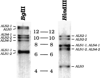
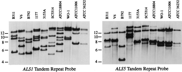
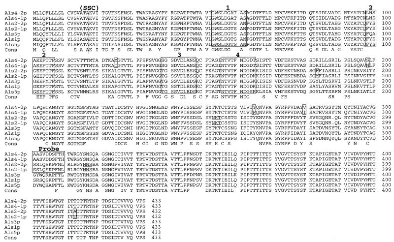
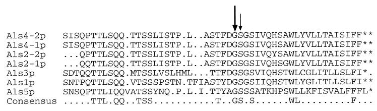
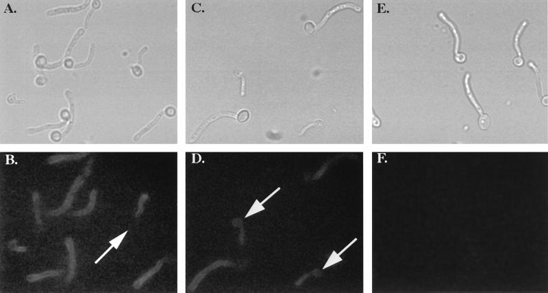
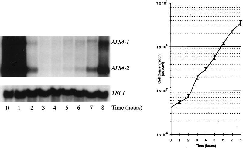
Additional genes in the growing ALS family of Candida albicans were isolated by PCR screening of a genomic fosmid library with primers designed from the consensus tandem-repeat sequence of ALS1. This procedure yielded fosmids encoding ALS2 and ALS4. ALS2 and ALS4 conformed to the three-domain structure of ALS genes, which consists of a central domain of tandemly repeated copies of a 108-bp motif, an upstream domain of highly conserved sequences, and a domain of divergent sequences 3' of the tandem repeats. Alignment of five predicted Als protein sequences indicated conservation of N- and C-terminal hydrophobic regions which have the hallmarks of secretory signal sequences and glycosylphosphatidylinositol addition sites, respectively. Heterologous expression of an N-terminal fragment of Als1p in Saccharomyces cerevisiae demonstrated function of the putative signal sequence with cleavage following Ala17. This signal sequence cleavage site was conserved in the four other Als proteins analyzed, suggesting identical processing of each protein. Primary-structure features of the five Als proteins suggested a cell-surface localization, which was confirmed by indirect immunofluorescence with an anti-Als antiserum. Staining was observed on mother yeasts and germ tubes, although the intensity of staining on the mother yeast decreased with elongation of the germ tube. Similar to other ALS genes, ALS2 and ALS4 were differentially regulated. ALS4 expression was correlated with the growth phase of the culture; ALS2 expression was not observed under many different in vitro growth conditions. The data presented here demonstrate that ALS genes encode cell-surface proteins and support the conclusion that the size and number of Als proteins on the C. albicans cell surface vary with strain and growth conditions.


Figures

FIG. 1
FIG. 1
Designation of genomic DNA fragments that encode ALS genes. Southern blots of BglII- or HindIII-digested genomic DNA from C. albicans 1161 were hybridized with an 870-bp KpnI fragment from ALS1 that contained only tandem-repeat sequences (26). ALS alleles corresponding to each of these restriction fragments were identified on separate blots with gene-specific probes developed previously (25, 26) or in this study. Allelic fragments were deduced by data from several experiments and sources, including physical mapping of C. albicans chromosomes (11, 48), restriction mapping of specific fosmid clones, DNA sequence analysis, and gene regulation studies (25, 26). Molecular size markers (in kilobases) are included between the blots.
FIG. 2
FIG. 2
Southern blots of BglII genomic DNA fragments hybridized with ALS1 and ALS5 tandem-repeat probes. Genomic DNA from a variety of C. albicans and C. stellatoidea strains was digested with BglII, blotted and probed with an 870-bp KpnI ALS1-tandem-repeat-specific probe (left panel). The blot was stripped and reprobed with a PCR-amplified ALS5 tandem-repeat fragment PCR amplified from C. albicans 1177 (right panel). Molecular size markers (in kilobases) are indicated at the left for each blot.
FIG. 3
FIG. 3
Alignment of N-terminal amino acid sequences predicted from genes in the ALS family. Amino acid sequences were predicted by the translation of ALS gene sequences 5′ of the start of the tandem-repeat domain. Sequences included are Als1p (26), Als2p and Als4p (this study), Als3p (25), and Als5p/Ala1p (18). Sequences were aligned with default parameters of the PILEUP program of Genetics Computer Group software (14). A consensus sequence (Cons), indicating amino acids conserved in all sequences, is provided below the alignment. The vertical line (designated SSC) between residues 17 and 18 denotes the site of signal sequence cleavage demonstrated biochemically for Als1p and predicted by computer algorithm to be conserved for the remaining proteins. Boxed regions labelled 1, 2, 3, and 4 correspond to the four 10-mer peptides from Als1p used to raise the rabbit polyclonal anti-Als antiserum used in indirect immunofluorescence studies. The boxed Als2p and Als4p sequences (labelled Probe) correspond to the region in the nucleotide sequence from which ALS2- and ALS4-specific oligonucleotides were derived. Boxed sequences between alleles of Als2p or Als4p indicate nonconserved amino acid sequences predicted from allelic nucleotide sequences. Consensus N glycosylation sites (2) are underlined in the Als2p sequences at positions 253 and 315. All CUG codons have been changed from Leu to Ser (46, 55).
FIG. 4
FIG. 4
Alignment of the C-terminal amino acid sequences of Als proteins. Approximately the last 50 amino acids of each predicted Als protein sequence were aligned, to demonstrate sequence conservation in this region. A consensus sequence indicating amino acids conserved in every protein is indicated below the multiple alignment. The putative GPI addition sites are indicated by arrows. The larger arrow over the Gly residue suggests that this is the more likely GPI attachment site.
FIG. 5
FIG. 5
Indirect immunofluorescence of C. albicans SC5314 germ tubes. YPD-grown C. albicans cells of strain SC5314 were induced to form germ tubes by inoculation into RPMI 1640 medium. Panels A, C, and E are light micrographs corresponding to the fluorescent micrographs in panels B, D, and F, respectively. Cells in panels B and D were treated with anti-Als antiserum followed by FITC-labelled goat-anti-rabbit IgG. Cells in panel F were stained with preimmune serum from the same rabbit in which the anti-Als serum was raised, followed by staining with the FITC-labelled secondary antiserum. The arrow in panel B indicates a large mother yeast cell that has lost its fluorescence. Arrows in panel D indicate small mother yeast cells for which fluorescence is still visible.
FIG. 6
FIG. 6
ALS4 expression in YPD-grown C. albicans cells. Strain SC5314 was grown overnight in YPD; cells from this culture were used to inoculate fresh YPD medium. Immediately after inoculation (0 h) and at each hour for the next 8 h, an aliquot of cells was removed. Cells were counted to generate a growth curve (right panel) and harvested for RNA extraction and Northern blotting with an ALS4-specific probe (left panel). A fragment of the C. albicans TEF1 gene was used as a control for equal loading of RNA.

References

    1. Aguiar J M, Baquero F, Jones J M. Candida albicans exocellular antigens released into a synthetic culture medium: characterization and serological response in rabbits. J Gen Microbiol. 1993;139:3005–3010. - PubMed
    1. Bause E. Structural requirements of N-glycosylation of proteins. Biochem J. 1983;209:331–336. - PMC - PubMed
    1. Bedell G W, Soll D R. Effects of low concentrations of zinc on the growth and dimorphism of Candida albicans: evidence for zinc-resistant and -sensitive pathways for mycelium formation. Infect Immun. 1979;26:348–354. - PMC - PubMed
    1. Brawner D L, Cutler J E. Ultrastructural and biochemical studies of two dynamically expressed cell surface determinants on Candida albicans. Infect Immun. 1986;51:327–336. - PMC - PubMed
    1. Brawner D L, Cutler J E. Variability in expression of a cell surface determinant on Candida albicans as evidenced by an agglutinating monoclonal antibody. Infect Immun. 1984;43:966–972. - PMC - PubMed

Publication types

MeSH terms

LinkOut - more resources