

Comparative Study
doi: 10.1128/JB.180.20.5448-5453.1998.
The 20S proteasome of Streptomyces coelicolor
Affiliations
- PMID: 9765579
- PMCID: PMC107596
- DOI: 10.1128/JB.180.20.5448-5453.1998
Item in Clipboard
Comparative Study
The 20S proteasome of Streptomyces coelicolor
J Bacteriol.
1998 Oct.
Abstract
20S proteasomes were purified from Streptomyces coelicolor A3(2) and shown to be built from one alpha-type subunit (PrcA) and one beta-type subunit (PrcB). The enzyme displayed chymotrypsin-like activity on synthetic substrates and was sensitive to peptide aldehyde and peptide vinyl sulfone inhibitors and to the Streptomyces metabolite lactacystin. Characterization of the structural genes revealed an operon-like gene organization (prcBA) similar to Rhodococcus and Mycobacterium spp. and showed that the beta subunit is encoded with a 53-amino-acid propeptide which is removed during proteasome assembly. The upstream DNA region contains the conserved orf7 and an AAA ATPase gene (arc).
Figures
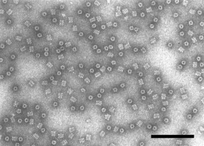
Electron micrograph of 20S proteasomes from S. coelicolor, negatively stained with uranyl acetate (3), showing ring-shaped end-on views representing projections along the cylinder axis and rectangular side views corresponding to projections perpendicular to the cylinder axis. Bar, 100 nm.
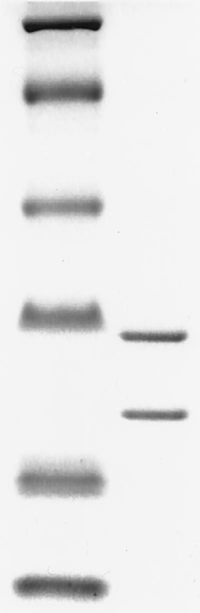
Tricine-SDS-polyacrylamide gel electrophoresis analysis of 2.28 μg of purified S. coelicolor 20S proteasomes (right lane). The sizes of the marker proteins (left lane) are 97.4, 66.2, 45.0, 31.0, 21.5, and 14.4 kDa (from top to bottom). The proteins were stained with Coomassie brilliant blue.
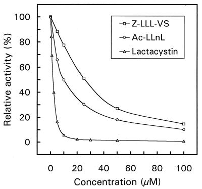
Inhibition of the S. coelicolor 20S proteasome by Ac-LLnL, Z-LLL-VS, and lactacystin. Purified proteasome (0.912 μg) was incubated with inhibitor in 100 μl of Tris-HCl (pH 8.0) at room temperature for 1 h. Subsequently, 20 nmol of Suc-LLVY-AMC was added. After incubation at 37°C for 1 h, the reaction was stopped (by addition of 100 μl of 10% SDS) and the amount of 7-amido-4-methylcoumarin released was measured.
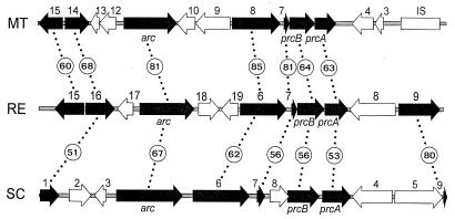
Gene organization of the proteasome structural genes (prcB, prcA) and the flanking regions from M. tuberculosis (MT) (GenBank accession no. Z97559; 14,030 bp), R. erythropolis (RE) (GenBank accession no. U26422 and this work; 13,000 bp), and S. coelicolor (SC) (this work; 10,612 bp). The fragment shown for Rhodococcus represents the second proteasome operon. The ORFs in the respective DNA regions are numbered independently. For M. tuberculosis, the ORF numbering of cosmid MTCY261 is used. The box labeled IS represents one of the sixteen copies of IS6110 present in the genome of M. tuberculosis H37Rv (8). Conserved ORFs and homologous genes are shown as filled arrows, and the extent of sequence conservation is indicated (numbers representing percentages of identical amino acids are circled).
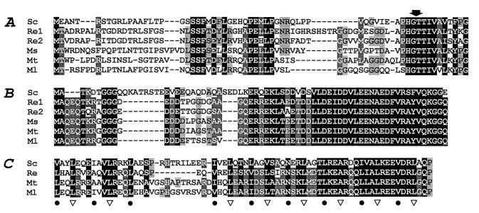
Multiple sequence alignments of the proteasome β-subunit propeptides (A), the ORF7 sequences (B), and the N-terminal coiled-coil regions of ARC homologues (C) from the following actinomycetes: M. leprae (Ml), M. smegmatis (Ms), M. tuberculosis (Mt), R. erythropolis (Re1 and Re2, or Re), and S. coelicolor (Sc). Differential shading (three levels in panels A and B; two levels in panel C) reflects the degree of sequence conservation (identical or similar amino acids) at a given position. The propeptide cleavage sites are marked with an arrow, and the first 10 residues of the processed β subunits are shown in panel A. In panel C, the hydrophobic amino acids in positions 1 and 4 of the heptad repeats of the proposed coiled-coil structures of ARC proteins are labeled • and ▿, respectively, and proline residues flanking coiled-coil segments are marked (in black on a shaded background).
References
-
- Akopian T N, Kisselev A F, Goldberg A L. Processive degradation of proteins and other catalytic properties of the proteasome from Thermoplasma acidophilum. J Biol Chem. 1997;272:1791–1798. - PubMed
-
- Baumeister W, Dahlmann B, Hegerl R, Kopp F, Kuehn L, Pfeifer G. Electron microscopy and image analysis of the multicatalytic proteinase. FEBS Lett. 1988;241:239–245. - PubMed
-
- Baumeister W, Walz J, Zühl F, Seemüller E. The proteasome: paradigm of a self-compartmentalizing protease. Cell. 1998;92:367–380. - PubMed
Publication types
MeSH terms
Substances
Associated data
- Actions
- Actions
LinkOut - more resources
Full Text Sources
Other Literature Sources
Molecular Biology Databases
Miscellaneous
