Comparative Study
. 1998 Oct;180(20):5478-83.
doi: 10.1128/JB.180.20.5478-5483.1998.

Gram-negative bacteria produce membrane vesicles which are capable of killing other bacteria

Affiliations
Comparative Study

Gram-negative bacteria produce membrane vesicles which are capable of killing other bacteria

Z Li et al. J Bacteriol. 1998 Oct.

Abstract

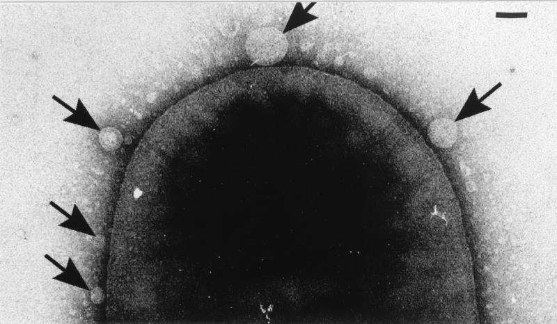
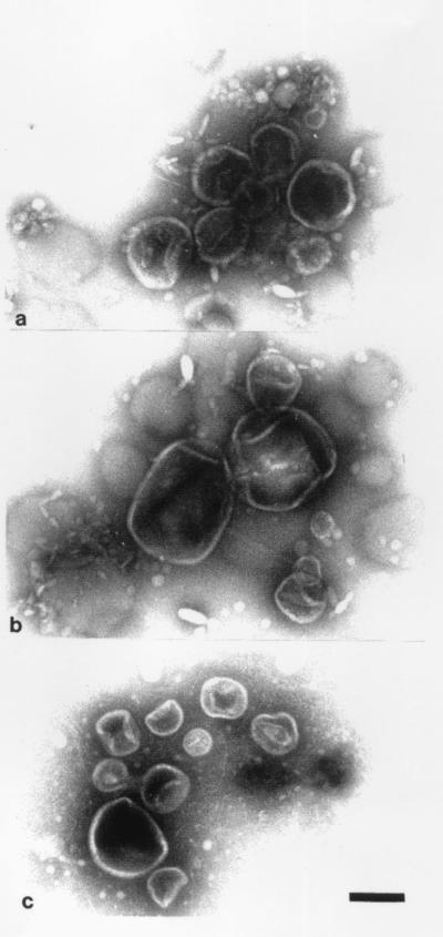
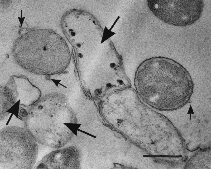
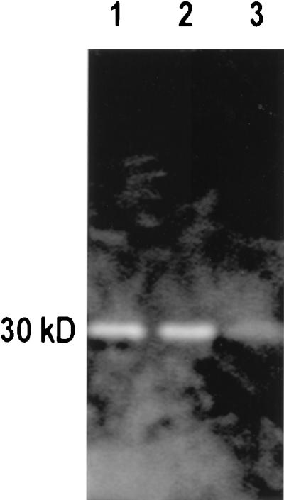
Naturally produced membrane vesicles (MVs), isolated from 15 strains of gram-negative bacteria (Citrobacter, Enterobacter, Escherichia, Klebsiella, Morganella, Proteus, Salmonella, and Shigella strains), lysed many gram-positive (including Mycobacterium) and gram-negative cultures. Peptidoglycan zymograms suggested that MVs contained peptidoglycan hydrolases, and electron microscopy revealed that the murein sacculi were digested, confirming a previous modus operandi (J. L. Kadurugamuwa and T. J. Beveridge, J. Bacteriol. 174:2767-2774, 1996). MV-sensitive bacteria possessed A1alpha, A4alpha, A1gamma, A2alpha, and A4gamma peptidoglycan chemotypes, whereas A3alpha, A3beta, A3gamma, A4beta, B1alpha, and B1beta chemotypes were not affected. Pseudomonas aeruginosa PAO1 vesicles possessed the most lytic activity.


Figures

FIG. 1
FIG. 1
Transmission electron micrograph of MVs forming on the surface of S. marcescens (arrows). The cell has been negatively stained with 2% (wt/vol) uranyl acetate and imaged in a Philips EM300 electron microscope operating under standard conditions at 60 kV. Bar = 100 nm.
FIG. 2
FIG. 2
MVs isolated from E. coli K-12 (a), S. marcescens (b), and P. aeruginosa PAO1 (c) which have been negatively stained and imaged as outlined in the legend to Fig. 1. All panels are the same magnification. Bar = 100 nm.
FIG. 3
FIG. 3
Cell of E. coli K-12 which was lysed by MVs from P. aeruginosa PAO1 during the agar plate assay. This preparation was fixed with 2% (wt/vol) glutaraldehyde followed by 2% (wt/vol) osmium tetroxide, embedded in LR white, and thin sectioned. The sections were stained with uranyl acetate and lead citrate and imaged as outlined in the legend to Fig. 1. The large arrows point to lysed cells, whereas the small arrows point to MVs that have the dimensions of those from P. aeruginosa. Bar = 1 μm.
FIG. 4
FIG. 4
Typical zymogram showing how peptidoglycan hydrolases can be separated by SDS-PAGE and identified by their digestion of murein sacculi within the gel. In this case, sacculi from E. coli K-12 are the substrates in the gel and the SDS-soluble components of MVs from S. marcescens UG-159 (lane 1), P. aeruginosa PAO1 (lane 2), and S. pullorum UG-147 (lane 3) have been run into the gel. Once separated from one another, the peptidoglycan hydrolases were renatured and the gels were stained according to Bernadsky et al. (1). All of the MVs from the three species possessed ∼30-kDa hydrolases that digested the E. coli murein sacculi.

References

    1. Bernadsky G, Beveridge T J, Clarke A J. Analysis of the sodium dodecyl sulfate-stable peptidoglycan autolysins of select gram-negative pathogens by using renaturing polyacrylamide gel electrophoresis. J Bacteriol. 1994;176:5225–5232. - PMC - PubMed
    1. Beveridge T J, Makin S A, Kadurugamuwa J L, Li Z S. Interactions between biofilms and the environment. FEMS Microbiol Rev. 1997;20:291–303. - PubMed
    1. Chatterjee S N, Das J. Electron microscopic observations on the excretion of cell-wall material by Vibrio cholerae. J Gen Microbiol. 1967;49:1–11. - PubMed
    1. Cleveland R F, Daneo-Moore L, Wicken A J, Shockman G D. Effect of lipoteichoic acid and lipids on lysis of intact cells of Streptococcus faecalis. J Bacteriol. 1976;127:1582–1584. - PMC - PubMed
    1. Cleveland R F, Höltje J-V, Wicken A J, Tomasz A, Daneo-Moore L, Shockman G D. Inhibition of bacterial wall lysis by lipoteichoic acids and related compounds. Biochem Biophys Res Commun. 1975;67:1128–1135. - PubMed

Publication types

MeSH terms

Substances

LinkOut - more resources