Review
. 1998 Oct;11(4):569-88.
doi: 10.1128/CMR.11.4.569.

Regulation and function of T-cell-mediated immunity during Toxoplasma gondii infection

Affiliations
Review

Regulation and function of T-cell-mediated immunity during Toxoplasma gondii infection

E Y Denkers et al. Clin Microbiol Rev. 1998 Oct.

Abstract

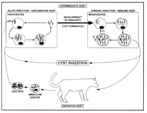
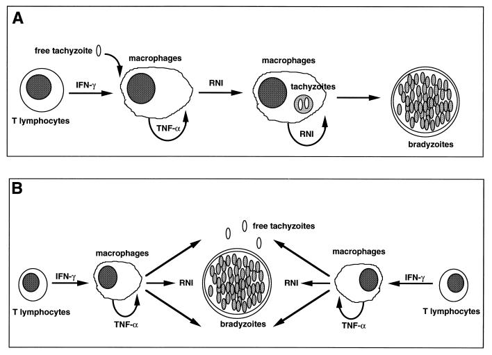
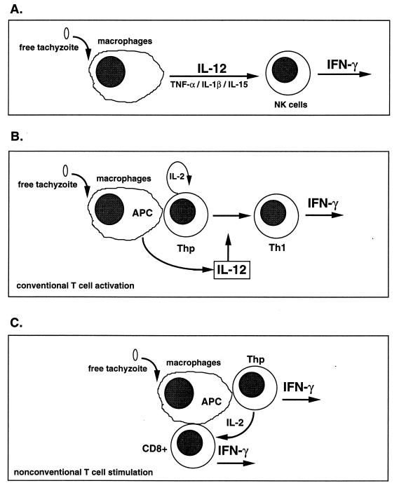
The intracellular protozoan Toxoplasma gondii is a widespread opportunistic parasite of humans and animals. Normally, T. gondii establishes itself within brain and skeletal muscle tissues, persisting for the life of the host. Initiating and sustaining strong T-cell-mediated immunity is crucial in preventing the emergence of T. gondii as a serious pathogen. The parasite induces high levels of gamma interferon (IFN-gamma) during initial infection as a result of early T-cell as well as natural killer (NK) cell activation. Induction of interleukin-12 by macrophages is a major mechanism driving early IFN-gamma synthesis. The latter cytokine, in addition to promoting the differentiation of Th1 effectors, is important in macrophage activation and acquisition of microbicidal functions, such as nitric oxide release. During chronic infection, parasite-specific T lymphocytes release high levels of IFN-gamma, which is required to prevent cyst reactivation. T-cell-mediated cytolytic activity against infected cells, while easily demonstrable, plays a secondary role to inflammatory cytokine production. While part of the clinical manifestations of toxoplasmosis results from direct tissue destruction by the parasite, inflammatory cytokine-mediated immunopathologic changes may also contribute to disease progression.


Figures

FIG. 1
FIG. 1
T. gondii life cycle. During acute infection, initiated by ingestion of cysts or oocysts, tachyzoites invade and proliferate within virtually any nucleated cell, resulting in host cell lysis and reinfection of more cells. Concurrent with the development of immunity, tachyzoites transform into slow-growing bradyzoites, which reside within cysts in tissues of the muscles and CNS. This chronic infection can persist for the life of the host, but in patients with immunodeficiency, cysts may rupture, leading to reinitiation of an acute infection. Within the gut of the cat, ingested parasites undergo differentiation into male and female gametes, which results in the formation of oocysts. These are shed in the feces and can remain infectious for many months.
FIG. 2
FIG. 2
(A) Cyst in an immunocompetent mouse chronically infected with T. gondii. (B and C) Satellite cysts (B) and clusters of free tachyzoites (arrows) (C) in the brains of chronically infected mice subsequently treated with anti-TNF-α or anti-IFN-γ MAb. These sections were stained with hematoxylin and eosin.
FIG. 3
FIG. 3
Alternative hypotheses for the control of T. gondii tachyzoite replication in tissues of immunocompetent hosts. (A) The cellular immune response plays an active role in driving encystment of the parasite. Recent evidence suggests that production of RNI may promote tachyzoite-bradyzoite transformation. (B) Initiation of cyst formation occurs independently of host immunity. Once cysts are established, a low rate of conversion of bradyzoites to tachyzoites occurs continuously, but these reemergent parasites are effectively controlled by components of type 1 cytokine-based immunity such as RNI.
FIG. 4
FIG. 4
Alternative pathways for induction of T-cell-independent and T-cell-dependent IFN-γ synthesis. (A) The T-cell-independent pathway is driven largely by IL-12, as well as TNF-α, IL-1β, and IL-15. These proinflammatory cytokines induce NK cell production of IFN-γ, which can promote macrophage activation and acquisition of microbiostatic activity, as well as Th1-cell differentiation. (B) Presentation of parasite peptide by a professional Ag-presenting cell (APC) such as a macrophage or dendritic cell to a Thp cell. This results in Th1 differentiation in the presence of T-cell-derived IL-2 and IL-12 produced by the Ag-presenting cell. (C) A less well-characterized but potentially very important pathway of early IFN-γ production, in which T. gondii is able to directly activate T cells in a manner that does not require IL-12.
FIG. 5
FIG. 5
Nonimmune CD4+ and CD8+ T lymphocytes proliferate in response to T. gondii and Ag-presenting cells. T lymphocytes from normal C57BL/6 mice (purified by passage over anti-mouse Ig columns) were subjected to treatment with rabbit complement and anti-CD4 MAb (to obtain CD8+ T cells) or anti-CD8 MAb (to obtain CD4+ T cells). The resulting populations were cultured with irradiated splenic adherent cells (SAC) that were either untreated, preincubated with T. gondii soluble Ag extract, or preinfected with RH strain tachyzoites. For some CD8+ populations, recombinant murine IL-2 was included. Proliferation was measured by incorporation of [3H]thymidine (3H-TdR), added 72 h after culture initiation.
FIG. 6
FIG. 6
Both β2-microglobulin and Aβ KO mice, which lack MHC class I-restricted CD8+ and class II-restricted CD4+ T lymphocytes, respectively, are susceptible to normally nonlethal T. gondii infection. Animals were infected by intraperitoneal injection of 20 cysts of the low-virulence ME49 parasite strain, and survival was monitored. Strain C57BL/6 mice (wild-type counterparts to the KO strains) do not begin to succumb to infection until approximately 100 days postinfection (48).
FIG. 7
FIG. 7
Dual role of CD8+ T lymphocytes as effectors of immunity to T. gondii. Priming of resting CD8+ cells, which requires parasite Ag, CD4+ T cells, and IL-2, results in anti-parasite effectors. These cells display MHC class I-restricted cytolytic activity toward Toxoplasma-infected target cells and release high levels of IFN-γ in response to parasite stimulation. Based upon studies in IFN-γ and perforin KO mouse strains, cytokine secretion is the major effector activity of CD8+ cells in acute and vaccine models of infection. IFN-γ acts, at least in part, through its ability to induce macrophage microbicidal functions such as NO production. Nevertheless, CTL function appears to play a secondary functional role in protection during chronic infection, as measured by increased mortality and incidence of brain cysts in perforin KO animals.

References

    1. Amichay D, Gazzinelli R T, Karupiah G, Moench T, Sher A, Farber J. The genes for the chemokines MuMig and Crg-2 are induced in protozoan and viral infections in response to IFN-γ with patterns of tissue expression that suggest nonredundant roles in vivo. J Immunol. 1996;157:4511–4520. - PubMed
    1. Anderson S E, Remington J S. Effect of normal and activated human macrophages on Toxoplasma gondii. J Exp Med. 1974;139:1154–1174. - PMC - PubMed
    1. Aosai F, Yang T-H, Ueda M, Yano A. Isolation of naturally processed peptides from a Toxoplasma gondii-infected human B lymphoma cell line that are recognized by cytotoxic T lymphocytes. J Parasitol. 1994;80:260–266. - PubMed
    1. Apt W B. Toxoplasmosis in developing countries. Parasitol Today. 1985;1:44–46. - PubMed
    1. Araujo F G. Depletion of CD4+ T cells but not inhibition of the protective activity of IFN-γ prevents cure of toxoplasmosis mediated by drug therapy in mice. J Immunol. 1992;149:3003–3007. - PubMed

Publication types