. 1998 Oct 13;95(21):12117-22.
doi: 10.1073/pnas.95.21.12117.

Inhibition of protease-resistant prion protein formation by porphyrins and phthalocyanines

Affiliations

Inhibition of protease-resistant prion protein formation by porphyrins and phthalocyanines

W S Caughey et al. Proc Natl Acad Sci U S A. .

Abstract

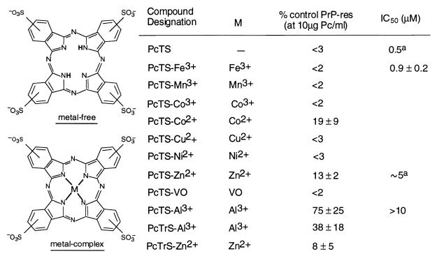
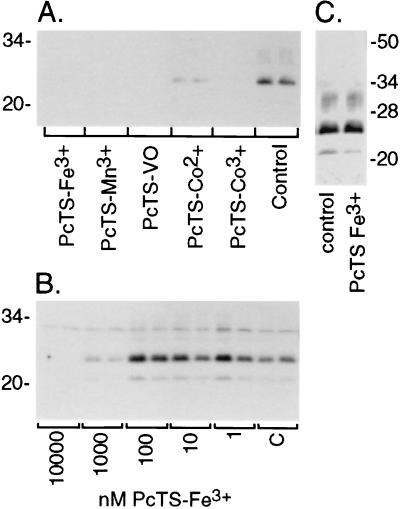
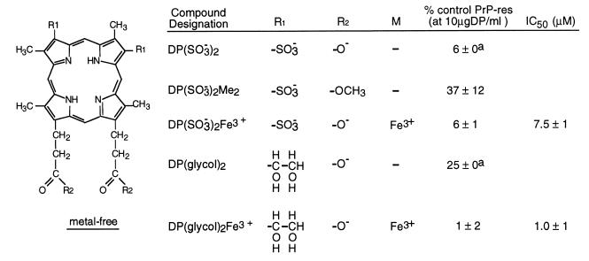
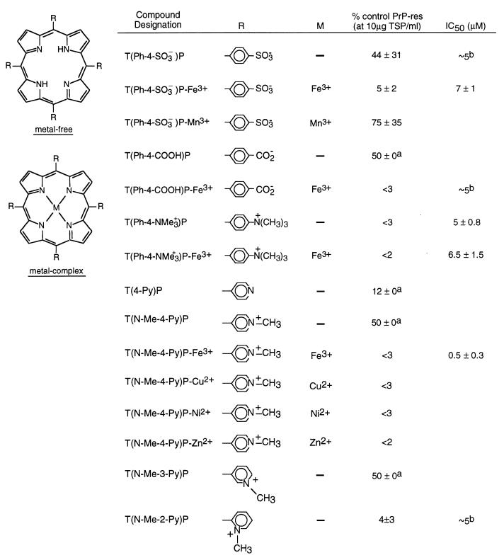
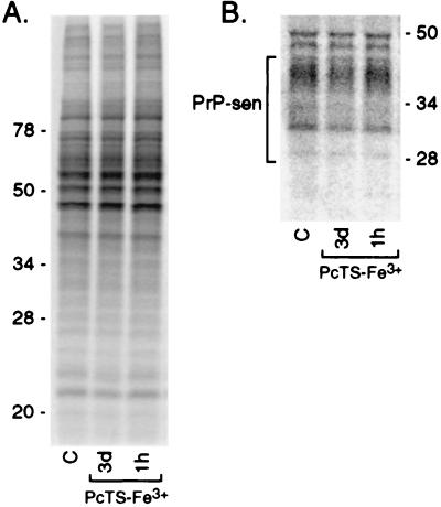
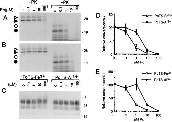
A central aspect of pathogenesis in the transmissible spongiform encephalopathies or prion diseases is the conversion of normal protease-sensitive prion protein (PrP-sen) to the abnormal protease-resistant form, PrP-res. Here we identify porphyrins and phthalocyanines as inhibitors of PrP-res accumulation. The most potent of these tetrapyrroles had IC50 values of 0.5-1 microM in scrapie-infected mouse neuroblastoma (ScNB) cell cultures. Inhibition was observed without effects on protein biosynthesis in general or PrP-sen biosynthesis in particular. Tetrapyrroles also inhibited PrP-res formation in a cell-free reaction composed predominantly of hamster PrP-res and PrP-sen. Inhibitors were found among phthalocyanines, deuteroporphyrins IX, and meso-substituted porphines; examples included compounds containing anionic, neutral protic, and cationic peripheral substituents and various metals. We conclude that certain tetrapyrroles specifically inhibit the conversion of PrP-sen to PrP-res without apparent cytotoxic effects. The inhibition observed in the cell-free conversion reaction suggests that the mechanism involved direct interactions of the tetrapyrrole with PrP-res and/or PrP-sen. These findings introduce a new class of inhibitors of PrP-res formation that represents a potential source of therapeutic agents for transmissible spongiform encephalopathies.


Figures

Figure 1
Figure 1
Inhibition of PrP-res formation in ScNB cells by phthalocyanine (Pc) sulfonates. ScNB cells were cultured for 4 days in the presence of Pcs as described in the text and analyzed for the accumulation of PrP-res by immunoblot (e.g., see Fig. 2). PrP-res band intensities are presented as mean percentage band intensity (±SD) relative to that from untreated control ScNB cells. All Pcs were tested at 10 μM and a few at lower concentrations to estimate the concentration giving 50% inhibition of PrP-res formation relative to control (IC50). PcTS and PcTrS designate compounds with four and three sulfonic acid groups, respectively, per molecule with only one on each peripheral, six-membered ring; variation in ring location results in a mixture of isomers. A superscript “a” indicates that a >80% drop in the 35S-PrP-res formation was observed between 10-fold dilutions of the inhibitor; we report the IC50 as the concentration halfway between the 10-fold dilutions tested, but the actual value could be ±50% of that value.
Figure 2
Figure 2
Immunoblots of inhibition of PrP-res accumulation in ScNB cultures by PcTS compounds. (A) Effects of PcTS compounds at 10 μM in the culture medium over 4 days. Control is without inhibitor. (B) Concentration dependence of effects of PcTS-Fe3+. “C” designates control. (C) Effect of treatment of ScNB cell lysates with 10 μM PcTS-Fe3+ for 1hr before PK treatment and extraction for the detection of PrP-res by immunoblot. For all of the immunoblots, the primary antibody R30 was used to identify PrP-res in the PK-digested cell extracts.
Figure 3
Figure 3
Inhibition of PrP-res formation in ScNB cells by sulfonate- and glycol-substituted deuteroporphyrins (DP). Analysis of effects of DPs on PrP-res accumulation was performed as described in the legend to Fig. 1 and Materials and Methods. A superscript “a” indicates that no difference in PrP-res immunoblot signal intensity could be discerned visually between replicates; however, with the autoradiographic methodology used, it was difficult to discriminate differences of ±5%.
Figure 4
Figure 4
Inhibition of PrP-res formation in ScNB cells by meso-tetrasubstituted porphines (TSP). Analysis of effects of TSPs on PrP-res accumulation was performed as described in the legend to Fig. 1 and Materials and Methods. A superscript “a” indicates that no difference in PrP-res immunoblot signal intensity could be discerned visually between replicates; however, with the autoradiographic methodology used, it was difficult to discriminate differences of ≈±5%. A superscript “b” indicates that a >80% drop in the 35S-PrP-res formation was observed between 10-fold dilutions of the inhibitor; we report the IC50 as the concentration halfway between the 10-fold dilutions tested, but the actual value could be ≈±50% of that value.
Figure 5
Figure 5
Phosphor autoradiographic images of 35S metabolic labeling of total proteins (A) and PrP-sen (B) in ScNB cells after pretreatments with 10 μM PcTS-Fe3+. Cultures were seeded and grown to confluence as done in experiments such as those presented in Fig. 2 A and B. PcTS-Fe3+ was added to the culture medium either 3 days or 1 h before labeling of the cells at confluence with [35S]methionine. PcTS-Fe3+ also was maintained at the same concentration in the labeling medium. In A, 5-μl aliquots of 1 ml cell lysates were run directly on the SDS/PAGE gel and the remainder of each lysate was used for the immunoprecipitation of the 35S-PrP-sen samples shown in B.
Figure 6
Figure 6
Inhibition of cell-free conversion of PrP-sen to PrP-res by PcTS-Fe3+ (A, D, and E) and PcTrS-Al3+ (B, D, and E) under GdnHCl-free (A, B, and D) or GdnHCl-containing conditions (E). 35S-PrP-sen was incubated with unlabeled PrP-res for 2 days in the presence of the designated concentration of phthalocyanine. One-tenth of the reaction was analyzed by SDS/PAGE without PK digestion; the remainder was digested with PK. (A and B) Phosphor autoradiographic images of 35S-PrP species; open and solid triangles, monoglycosylated and unglycosylated 35S-PrP, respectively, without PK treatment; open and solid circles, monoglycosylated and unglycosylated 35S-PrP-res, respectively, after PK digestion. (C) Immunoblot analysis of the total PrP-res in the PK-digested reaction products using mAb 3F4 [which has an epitope within the normally PK-resistant portion of PrP-res (50)] as described (51). Molecular mass markers are designated in kDa along the right side of AC. The loss of 35S-PrP in the 100 μM PcTS-Fe3+ lane (-PK) appeared to be due to SDS-insoluble aggregation because higher-molecular-mass 35S-PrP species were visible near the top of the lane (not shown). Because this apparent aggregation was not observed with lower, but highly inhibitory, concentrations of PcTS-Fe3+ (e.g., 1 μM) or with PcTrS-Al3+ or several other inhibitory tetrapyrroles described in the text, we conclude that it was not related to inhibition. (D and E) Graphs of the quantitated 35S-PrP-res products (bands marked with circles in A and B) using GdnHCl-free or GdnHCl-containing conditions, respectively. The data points show the mean ± SD of triplicate determinations.

References

    1. Caughey B, Chesebro B. Trends Cell Biol. 1997;7:56–62. - PubMed
    1. Caughey B, Race R E. J Neurochem. 1992;59:768–771. - PubMed
    1. Caughey B, Raymond G J. J Virol. 1993;67:643–650. - PMC - PubMed
    1. Caughey B, Ernst D, Race R E. J Virol. 1993;67:6270–6272. - PMC - PubMed
    1. Ehlers B, Diringer H. J Gen Virol. 1984;65:1325–1330. - PubMed