. 1998 Oct 13;95(21):12153-7.
doi: 10.1073/pnas.95.21.12153.

The anti-angiogenic agent fumagillin covalently modifies a conserved active-site histidine in the Escherichia coli methionine aminopeptidase

Affiliations

The anti-angiogenic agent fumagillin covalently modifies a conserved active-site histidine in the Escherichia coli methionine aminopeptidase

W T Lowther et al. Proc Natl Acad Sci U S A. .

Abstract

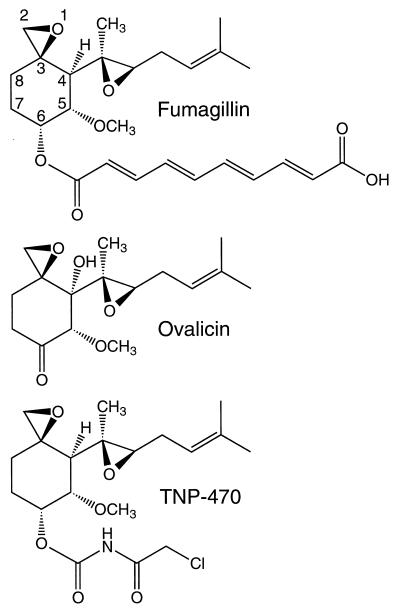
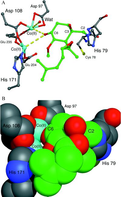
Methionine aminopeptidase (MetAP) exists in two forms (type I and type II), both of which remove the N-terminal methionine from proteins. It previously has been shown that the type II enzyme is the molecular target of fumagillin and ovalicin, two epoxide-containing natural products that inhibit angiogenesis and suppress tumor growth. By using mass spectrometry, N-terminal sequence analysis, and electronic absorption spectroscopy we show that fumagillin and ovalicin covalently modify a conserved histidine residue in the active site of the MetAP from Escherichia coli, a type I enzyme. Because all of the key active site residues are conserved, it is likely that a similar modification occurs in the type II enzymes. This modification, by occluding the active site, may prevent the action of MetAP on proteins or peptides involved in angiogenesis. In addition, the results suggest that these compounds may be effective pharmacological agents against pathogenic and resistant forms of E. coli and other microorganisms.


Figures

Figure 1
Figure 1
The anti-angiogenesis compounds fumagillin, ovalicin, and TNP-470. The intact epoxide attached to C3 is required for anti-angiogenic activity. Numbering scheme taken from Griffith et al. (14).
Figure 2
Figure 2
Analysis of the MetAP-fum complex. (A) Electronic absorption spectrum of E. coli MetAP before (solid line) and after (dashed line) fumagillin treatment and removal of excess fumagillin. The peak at 280 nm derives from the protein aromatic residues whereas the peak at 335 nm matches the absorbance of the conjugated fumagillin double-bond system. The absorption spectrum of the MetAP-ovalicin complex completely lacked the latter peak (not shown). (B) RP-HPLC separation monitored at 220 nm of the MetAP-fum covalent complex after digestion with Endo Glu-C and Endo Lys-C, and treatment with iodoacetic acid. Simultaneous monitoring of the region at 53 min (see arrow) at 335 nm (Inset), uniquely identified the fumagillin-labeled peak (∗) in the complex peptide mixture.
Figure 3
Figure 3
Mass spectral analysis of the isolated, fumagillin-linked peptide. (A) A subset of the observed mass fragmentation spectra of the MetAP 76–86 polypeptide containing the fumagillin covalent adduct. The m/z ions are the result of cleavage at the peptide bonds. Fragments with m/z 147.2, 199.2, 1338.8, 1453.6, and 1524.8 values (not shown) also were observed and assigned to the theoretical fragmentation pattern shown in B. Peak heights are normalized to m/z 218. (B) Summary of the theoretical peptide fragmentation pattern with corresponding molecular weights. The spectrum in A is consistent with the addition of fumagillin to His-79 (FumH) and the alkylation of Cys-78 (CMC).
Figure 4
Figure 4
Electronic absorption spectra from the d→d transitions of the Co(II) ions in the active site of E. coli MetAP (solid line), and the MetAP-fum (long dashed line) and MetAP-ovalicin (short dashed line) covalent complexes. The molar absorptivities were calculated with respect to protein concentration.
Figure 5
Figure 5
Hypothetical model of ovalicin bound covalently to His-79 in the active site of E. coli MetAP. (A) An energy-minimized model of the complex was made assuming sp3 atoms at C2 and C3 after formation of the covalent bond between C2 and Nɛ2 of His-79. The His-79-ovalicin complex was rotated about the His-79 Cα-Cβ and Cβ-Cγ bonds until the C6 keto oxygen atom of ovalicin was within approximately 2.5 Å of each Co(II) ion. As illustrated via a space filling model (B), access to the metal center is almost completely occluded. Color scheme: protein carbon atoms (gray), ovalicin carbon atoms (green), nitrogen (blue), oxygen (red), cobalt (cyan), and sulfur (yellow). Energy minimizations were performed by using the discover program in the InsightII package (Biosym Technologies, San Diego/Molecular Simulations, Waltham, MA). The figure was made by using midasplus (University of California, San Francisco Computer Graphics Laboratory).

References

    1. Ingber D, Fujita T, Kishimoto S, Sudo K, Kanamaru T, Brem H, Folkman J. Nature (London) 1990;348:555–557. - PubMed
    1. Corey E J, Guzman-Perez A, Noe M C. J Am Chem Soc. 1994;116:12109–12110.
    1. O’Reilly M S, Boehm T, Shing Y, Fukai N, Vasios G, Lane W S, Flynn E, Birkhead J R, Olsen B R, Folkman J. Cell. 1997;88:277–285. - PubMed
    1. O’Reilly M S, Holmgren L, Shing Y, Chen C, Rosenthal R A, Moses M, Lane W S, Cao Y, Sage E H, Folkman J. Cell. 1994;79:315–328. - PubMed
    1. Zetter B R. Annu Rev Med. 1998;49:407–424. - PubMed

Publication types

LinkOut - more resources