

The helical domain of intestinal fatty acid binding protein is critical for collisional transfer of fatty acids to phospholipid membranes
- PMID: 9770459
- PMCID: PMC22804
- DOI: 10.1073/pnas.95.21.12174
The helical domain of intestinal fatty acid binding protein is critical for collisional transfer of fatty acids to phospholipid membranes
Abstract
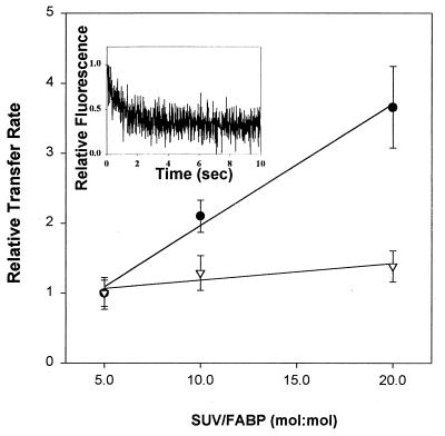
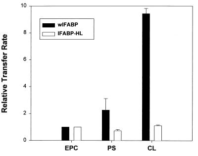
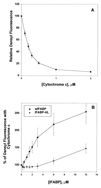
Fatty acid binding proteins (FABPs) exhibit a beta-barrel topology, comprising 10 antiparallel beta-sheets capped by two short alpha-helical segments. Previous studies suggested that fatty acid transfer from several FABPs occurs during interaction between the protein and the acceptor membrane, and that the helical domain of the FABPs plays an important role in this process. In this study, we employed a helix-less variant of intestinal FABP (IFABP-HL) and examined the rate and mechanism of transfer of fluorescent anthroyloxy fatty acids (AOFA) from this protein to model membranes in comparison to the wild type (wIFABP). In marked contrast to wIFABP, IFABP-HL does not show significant modification of the AOFA transfer rate as a function of either the concentration or the composition of the acceptor membranes. These results suggest that the transfer of fatty acids from IFABP-HL occurs by an aqueous diffusion-mediated process, i.e., in the absence of the helical domain, effective collisional transfer of fatty acids to membranes does not occur. Binding of wIFABP and IFABP-HL to membranes was directly analyzed by using a cytochrome c competition assay, and it was shown that IFABP-HL was 80% less efficient in preventing cytochrome c from binding to membranes than the native IFABP. Collectively, these results indicate that the alpha-helical region of IFABP is involved in membrane interactions and thus plays a critical role in the collisional mechanism of fatty acid transfer from IFABP to phospholipid membranes.
Figures
References
-
- Banaszak L, Winter N, Xu Z, Bernlohr D A, Cowan S, Jones T A. Adv Protein Chem. 1994;45:89–151. - PubMed
-
- Veerkamp J H, Maatman R G. Prog Lipid Res. 1995;34:17–52. - PubMed
-
- Sacchetini J C, Gordon J I. J Biol Chem. 1993;268:18399–18402. - PubMed
-
- Sacchettini J C, Gordon J I, Banaszak L. J Mol Biol. 1989;208:327–339. - PubMed
-
- Hodsdon M E, Cistola D P. Biochemistry. 1997;36:2278–2290. - PubMed
Publication types
MeSH terms
Substances
Grants and funding
LinkOut - more resources
Full Text Sources
