. 1998 Oct 13;95(21):12202-7.
doi: 10.1073/pnas.95.21.12202.

Rsk-2 activity is necessary for epidermal growth factor-induced phosphorylation of CREB protein and transcription of c-fos gene

Affiliations

Rsk-2 activity is necessary for epidermal growth factor-induced phosphorylation of CREB protein and transcription of c-fos gene

D De Cesare et al. Proc Natl Acad Sci U S A. .

Abstract

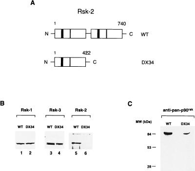
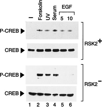
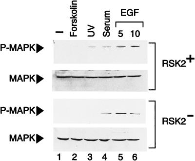
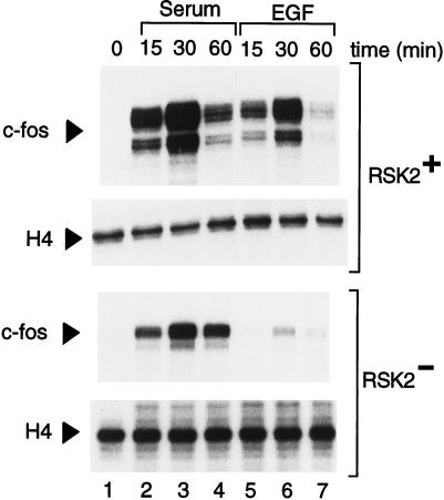
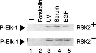
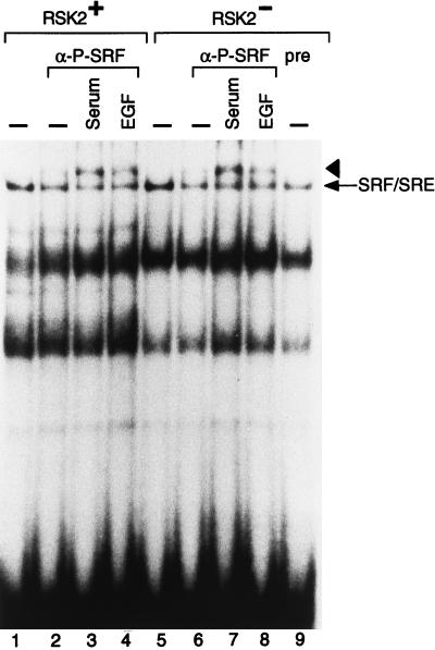
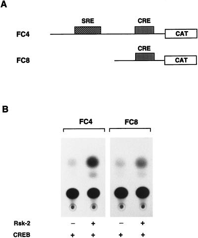
Activation by growth factors of the Ras-dependent signaling cascade results in the induction of p90 ribosomal S6 kinases (p90(rsk)). These are translocated into the nucleus upon phosphorylation by mitogen-activated protein kinases, with which p90(rsk) are physically associated in the cytoplasm. In humans there are three isoforms of the p90(rsk) family, Rsk-1, Rsk-2, and Rsk-3, which are products of distinct genes. Although these isoforms are structurally very similar, little is known about their functional specificity. Recently, mutations in the Rsk-2 gene have been associated with the Coffin-Lowry syndrome (CLS). We have studied a fibroblast cell line established from a CLS patient that bears a nonfunctional Rsk-2. Here we document that in CLS fibroblasts there is a drastic attenuation in the induced Ser-133 phosphorylation of transcription factor CREB (cAMP response element-binding protein) in response to epidermal growth factor stimulation. The effect is specific, since response to serum, cAMP, and UV light is unaltered. Furthermore, epidermal growth factor-induced expression of c-fos is severely impaired in CLS fibroblasts despite normal phosphorylation of serum response factor and Elk-1. Finally, coexpression of Rsk-2 in transfected cells results in the activation of the c-fos promoter via the cAMP-responsive element. Thus, we establish a link in the transduction of a specific growth factor signal to changes in gene expression via the phosphorylation of CREB by Rsk-2.


Figures

Figure 1
Figure 1
Rsk-2 mutation in DX34 Coffin–Lowry patient. (A Upper) Schematic representation of the wild-type Rsk-2 protein. (Lower) Predicted Rsk-2 protein in fibroblasts from DX34 Coffin–Lowry patient (14). Numbers refer to the amino acid length of the proteins. ATP-binding sites are shown as solid boxes, and APE (A = alanine, P = proline, E = glutamic acid) sites are shown as hatched boxes. (B) Levels of Rsk-1, Rsk-3, and Rsk-2 in normal and CLS fibroblasts. Total cell lysates from cells were analyzed by Western blot by using anti-Rsk-1, Rsk-3, and Rsk-2 specific antibodies (see Materials and Methods). Rsk-2 is not detectable in DX34 cells as the antibody is directed against the C terminus of the protein. (C) Levels of p90rsk using a general anti-p90rsk antibody (see Materials and Methods). The total level of p90rsk is lower in DX34 cells as the contribution of Rsk-2 is missing. There is no evidence for a truncated Rsk-2 protein in DX34 cells.
Figure 2
Figure 2
Rsk-2 is necessary for CREB phosphorylation upon activation of EGF signaling pathway. Normal (RSK2+) and CLS (RSK2) cells were starved 24 hr in the absence of serum before harvesting. Cell extracts were quantified by Coomassie blue staining. Lanes: 2, forskolin was utilized at a concentration of 10 μg/ml for 30-min stimulation; 3, cells were irradiated for 10 sec and harvested 30 min after exposure to UV (as in ref. 20); 4, fetal calf serum was added at 20% final concentration for 10 min; 5 and 6, cells were exposed to 30 ng/ml of EGF for 5 and 10 min (lanes 5 and 6, respectively). CREB phosphorylation levels in Rsk-2 cells upon EGF induction are about 5% of the forskolin-induced level in the same cells and 10% of the corresponding EGF induction in Rsk-2+ cells, after adjusting with the basal levels.
Figure 3
Figure 3
MAPK response is normal in cells from CLS patients. Normal (RSK2+) and CLS (RSK2) cells were starved 24 hr in the absence of serum before treatment and then harvested. An antibody recognizing the phosphorylated MAPK was used (see Materials and Methods). Lanes: 2, forskolin was added at the concentration of 10 μg/ml for 10 min; 3, cells were exposed for 10 sec to UV rays and harvested 30 min after irradiation (20); 4, samples were stimulated in the presence of 20% fetal calf serum for 10 min; 5 and 6, cells were treated with 30 ng/ml of EGF for the indicated times. Total levels of MAPK are uniform in all samples.
Figure 4
Figure 4
Induction of c-fos transcription by EGF requires Rsk-2 activity. Normal (RSK2+) and CLS (RSK2) fibroblasts were starved 24 hr before induction with serum and EGF and then harvested at the indicated times. Ten micrograms of total RNA was used per lane, and RNase protection was performed as described (see Materials and Methods). Lanes: 2–4, cells were stimulated with 20% fetal calf serum; 5–7, fibroblasts were exposed to 30 ng/ml of EGF. The peak of c-fos-induced expression by EGF in the Rsk-2 cells is 5% of the signal obtained with serum in the same cells and 2% of the EGF peak in the Rsk-2+ cells (after adjusting with basal levels). Transcript levels of histone H4 were monitored as internal control.
Figure 5
Figure 5
Rsk-2 is not necessary for TCF/Elk-1 phosphorylation. Normal (RSK2+) and CLS (RSK2) fibroblasts were deprived of serum for 24 hr before treatment. Lanes: 2, 10 μg/ml of forskolin were used for 30-min stimulation; 3, cells were UV-irradiated for 10 sec and harvested 20 min later (19); 4, fetal calf serum was added at a 20% concentration; 5, EGF was used at the concentration of 30 ng/ml for 10-min stimulation.
Figure 6
Figure 6
Rsk-2 is not necessary for SRF phosphorylation. Normal (RSK2+) and CLS (RSK2) cells that had been starved for 24 hr were treated with 20% fetal calf serum and 30 ng/ml of EGF for 10 min before preparing nuclear extracts. Three micrograms of nuclear proteins was used per lane. α-Phospho-SRF (α-P-SRF) antibodies were added to the equilibrium-binding reactions (lanes 3–5 and 6–8) for 10 min. The specific SRF/SRE complex is indicated by an arrow. The SRF/SRE/α-P-SRF super-shifted complex is indicated by an arrowhead. Lane 9: equilibrium-binding reaction was incubated with preimmune serum for 10 min before loading onto the polyacrylamide gel.
Figure 7
Figure 7
The CRE is sufficient to mediate induction of c-fos expression by Rsk-2. (A) Schematic representation of FC4 and FC8 reporter plasmids (35); binding sites for Elk-1/SRF (SRE) and CREB (CRE) are indicated. (B) COS cells were transfected with 0.5 μg of FC4 and FC8 reporter plasmids in combination with 0.2 μg of CREB expression vector and 2 μg of Rsk-2 expression vector when indicated. Cells were harvested 36 hr after addition of calcium phosphate precipitate. Rsk-2-mediated activation is 7.5 (±0.5)-fold for FC4 and 4.5 (±0.5)-fold for FC8, as established by photometric scanning of the autoradiograms. The results presented are representative of three independent experiments.

References

    1. Qiu M S, Green S H. Neuron. 1991;7:937–946. - PubMed
    1. Thomas S M, De Marco M, D’Arcangelo G, Halegoua S, Brugge J S. Cell. 1992;68:1031–1040. - PubMed
    1. Treisman R. Curr Opin Cell Biol. 1996;8:205–215. - PubMed
    1. Robinson M J, Cobb M H. Curr Opin Cell Biol. 1997;9:180–186. - PubMed
    1. Blenis J. Proc Natl Acad Sci USA. 1993;90:5889–5892. - PMC - PubMed

Publication types

MeSH terms

LinkOut - more resources