. 1998 Oct 13;95(21):12208-13.
doi: 10.1073/pnas.95.21.12208.

Identification of a human nuclear receptor defines a new signaling pathway for CYP3A induction

Affiliations

Identification of a human nuclear receptor defines a new signaling pathway for CYP3A induction

G Bertilsson et al. Proc Natl Acad Sci U S A. .

Abstract

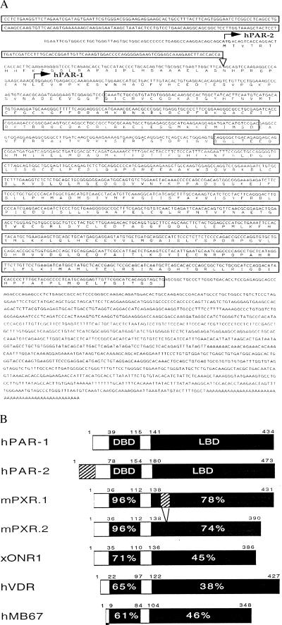
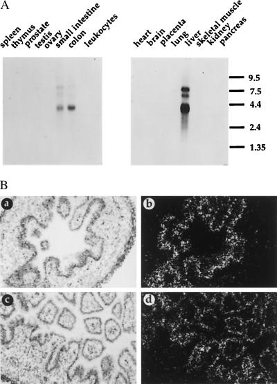
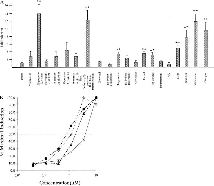
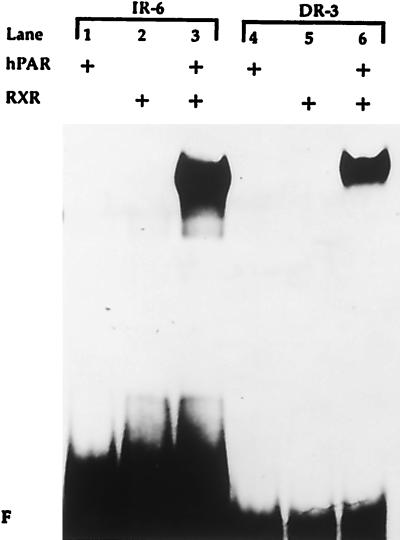
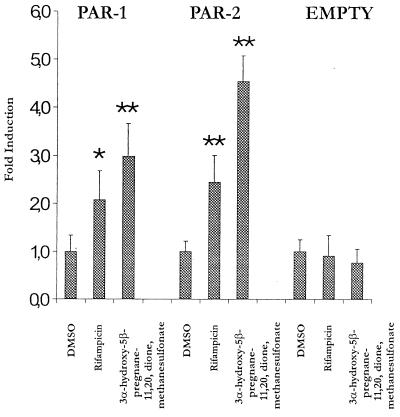
Nuclear receptors regulate metabolic pathways in response to changes in the environment by appropriate alterations in gene expression of key metabolic enzymes. Here, a computational search approach based on iteratively built hidden Markov models of nuclear receptors was used to identify a human nuclear receptor, termed hPAR, that is expressed in liver and intestines. hPAR was found to be efficiently activated by pregnanes and by clinically used drugs including rifampicin, an antibiotic known to selectively induce human but not murine CYP3A expression. The CYP3A drug-metabolizing enzymes are expressed in gut and liver in response to environmental chemicals and clinically used drugs. Interestingly, hPAR is not activated by pregnenolone 16alpha-carbonitrile, which is a potent inducer of murine CYP3A genes and an activator of the mouse receptor PXR.1. Furthermore, hPAR was found to bind to and trans-activate through a conserved regulatory sequence present in human but not murine CYP3A genes. These results provide evidence that hPAR and PXR.1 may represent orthologous genes from different species that have evolved to regulate overlapping target genes in response to pharmacologically distinct CYP3A activators, and have potential implications for the in vitro identification of drug interactions important to humans.


Figures

Figure 1
Figure 1
hPAR is a novel member of the nuclear receptor family. (A) Nucleotide and predicted amino acid sequence of hPAR-1 and -2. The putative initiation codons for hPAR-1 and -2 are indicated by solid arrows. The 5′ untranslated region of hPAR-1 is boxed until the splice site indicated by an open arrow. The predicted LBD and DBD are boxed. (B) Amino acid sequence comparison between hPAR and related nuclear receptors. The similarity in the DBD and LBD between hPAR and related nuclear receptors is indicated as percentage amino acid identity. The N-terminal region in hPAR-2 that is different from hPAR-1 is indicated by the cross-hatched box in hPAR-2.
Figure 2
Figure 2
Expression pattern of hPAR. (A) Northern blot analysis of adult human tissues. RNA size markers are indicated to the right in kb. (B) In situ analysis of embryonic hPAR mRNA expression. Bright- (a and c) and dark-field (b and d) views of hPAR expression in the intestine of a 10-week-old human embryo are shown. Specificity was determined by using sense-probe controls to adjacent tissue sections as the antisense probe. No signal could be detected with the sense probe.
Figure 3
Figure 3
Activation profile of hPAR in transiently transfected Caco-2 cells. (A) Caco-2 cells were transfected with luciferase reporter plasmid and expression plasmid encoding GAL4-hPAR chimeric protein. Cells then were treated with vehicle (DMSO) or 10 μM of the indicated compounds for 24 hr. Cell extracts were analyzed for luciferase activity, and data represent the mean ± SD. ∗∗, P < 0.001 (Student’s t test) as compared with the DMSO control. (B) Caco-2 cells were transfected with luciferase reporter plasmid and expression plasmid encoding GAL4-hPAR chimeric protein. Cells then were treated with the indicated concentration of clotrimazole (solid circles), 3α-hydroxy-5β-pregnane-11,20,dione, methanesulfonate (open diamonds), Rifampicine (solid triangles), or Nifidipine (asterisks) for 24 hr. Cell extracts were analyzed for luciferase activity, and data are plotted as the percentage of maximal induction.
Figure 4
Figure 4
DNA binding of hPAR to conserved human CYP3A regulatory element. Gel mobility-shift assay. In vitro translated hPAR-1 was incubated in the presence or absence of in vitro translated RXRb together with radiolabeled oligonucleotides containing either the CYP3A sequence (IR-6) or a direct repeat separated by 3 nt (DR-3) as indicated.
Figure 5
Figure 5
Activation of hPAR through the CYP3A4 IR-6 element. Caco-2 cells were transfected with the CYP3A4 IR-6 luciferase reporter plasmid with (PAR-1 and PAR-2) or without (EMPTY) expression plasmid encoding either hPAR-1 or hPAR-2 protein as indicated. The cells were treated with vehicle (DMSO) or 10 μM of the indicated compounds for 24 hr. Cell extracts were analyzed for luciferase activity, and data represent the mean ± SD. ∗∗, P < 0.001; ∗, P < 0.05 (Student’s t test) as compared with the DMSO controls.

References

    1. Altschul S F, Boguski M S, Gish W, Wooton J C. Nat Genet. 1994;6:119–129. - PubMed
    1. Krogh A, Brown M, Mian I S, Sjölander K, Haussler D. J Mol Biol. 1994;235:1501–1531. - PubMed
    1. Baldi P, Chauvin Y, Hunkapiller T, McClure M A. Proc Natl Acad Sci USA. 1994;91:1059–1063. - PMC - PubMed
    1. Escriva H, Safi R, Hänni C, Langlois M-C, Saumito-Laprade P, Stehelin D, Capron A, Pierce R, Laudet V. Proc Natl Acad Sci USA. 1997;94:6803–6808. - PMC - PubMed
    1. Mangelsdof D J, Thummel C, Beato M, Herrlich P, Schütz G, Umesono K, Blumberg B, Kastner P, Mark M, Chambon P, Evans R M. Cell. 1995;83:835–839. - PMC - PubMed

Publication types

MeSH terms

Associated data