. 1998 Oct 13;95(21):12226-31.
doi: 10.1073/pnas.95.21.12226.

Mammalian capping enzyme binds RNA and uses protein tyrosine phosphatase mechanism

Affiliations

Mammalian capping enzyme binds RNA and uses protein tyrosine phosphatase mechanism

Y Wen et al. Proc Natl Acad Sci U S A. .

Abstract

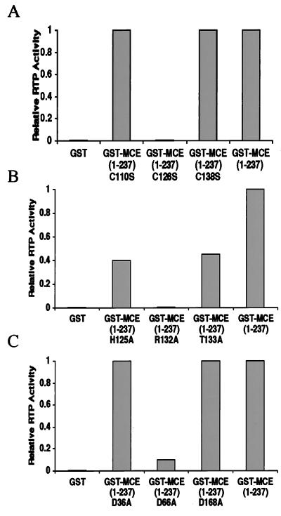
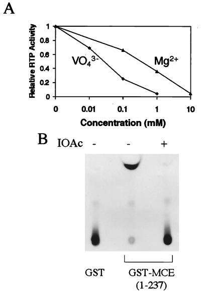
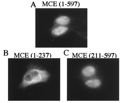
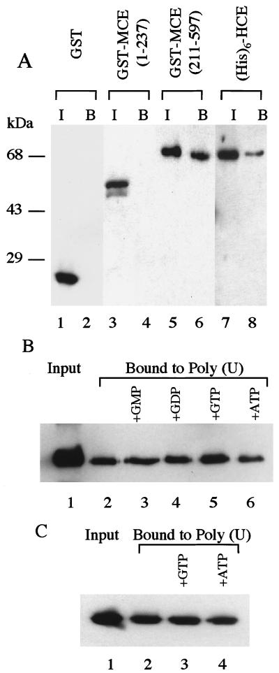
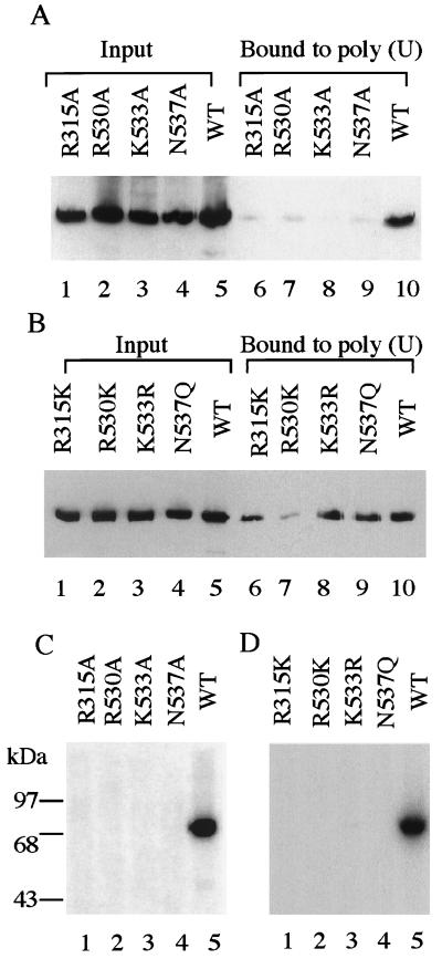
Mammalian capping enzymes are bifunctional proteins with both RNA 5'-triphosphatase and guanylyltransferase activities. The N-terminal 237-aa triphosphatase domain contains (I/V)HCXXGXXR(S/T)G, a sequence corresponding to the conserved active-site motif in protein tyrosine phosphatases (PTPs). Analysis of point mutants of mouse RNA 5'-triphosphatase identified the motif Cys and Arg residues and an upstream Asp as required for activity. Like PTPs, this enzyme was inhibited by iodoacetate and VO43- and independent of Mg2+, providing additional evidence for phosphate removal from RNA 5' ends by a PTP-like mechanism. The full-length, 597-aa mouse capping enzyme and the C-terminal guanylyltransferase fragment (residues 211-597), unlike the triphosphatase domain, bound poly (U) and were nuclear in transfected cells. RNA binding was increased by GTP, and a guanylylation-defective, active-site mutant was not affected. Ala substitution at positions required for the formation of the enzyme-GMP capping intermediate (R315, R530, K533, or N537) also eliminated poly (U) binding, while proteins with conservative substitutions at these sites retained binding but not guanylyltransferase activity. These results demonstrate that the guanylyltransferase domain of mammalian capping enzyme specifies nuclear localization and RNA binding. Association of capping enzyme with nascent transcripts may act in synergy with RNA polymerase II binding to ensure 5' cap formation.


Figures

Figure 1
Figure 1
C126, R132, H125, T133, and D66 are important for mouse RTP activity. γ-32P-labeled RNA was incubated with 100 ng of the indicated purified proteins as described in Materials and Methods. Released 32Pi was quantitated by PhosphorImager. Values are expressed relative to 1.0 for GST-MCE (1–237). Variation among several experiments was ±0.1.
Figure 2
Figure 2
VO43−, Mg2+, and IOAc inhibit mouse RTP activity. Purified GST-MCE (1–237) was assayed for RTP activity as in Fig. 1 in the presence of the indicated concentrations of Na3VO4 or MgCl2 (A) or IOAc (B).
Figure 3
Figure 3
Full-length capping enzyme and C-terminal guanylyltransferase fragment are localized in nuclei. 3T3 cells transfected with pcB6+2myc containing MCE (1–597) (A), (1–237) (B), or (211–597) (C) were analyzed by immunofluorescence staining as described in Materials and Methods.
Figure 4
Figure 4
Full-length capping enzyme and C-terminal guanylyltransferase fragment bind poly (U). (A) Purified GST, GST-MCE (1–237), GST-MCE (211–597), and (His)6-HCE were incubated with poly (U) beads as described in Materials and Methods. Bound proteins (lanes 2, 4, 6, and 8) were analyzed by SDS/PAGE and Western blot in comparison with the input (lanes 1, 3, 5, and 7). Immunoblots were developed by anti-GST antibody (lanes 1–6) or anti-MCE antibody (lanes 7 and 8). (B) Purified GST-MCE (211–597) (lane 1) was incubated with poly (U) beads (lane 2) in the presence of 30 μM each of GMP (lane 3), GDP (lane 4), GTP (lane 5), or ATP (lane 6). Bound proteins were analyzed by SDS/PAGE and Western blot analysis with anti-MCE antibody. (C) Purified (His)6-MCE (211–597) K294A (lane 1) was incubated with poly (U) beads (lane 2) and 30 μM GTP (lane 3) or ATP (lane 4) followed by analysis as in B.
Figure 5
Figure 5
R315, R530, K533, and N537 are involved in both poly (U) binding and guanylylation. (A and B) Purified GST-MCE (211–597) (lane 5) and the indicated mutants (lanes 1–4) were incubated with poly (U) beads, and bound proteins (lanes 6–10) were analyzed in comparison with the input as in Fig. 4A. (C and D) Purified GST-MCE (211–597) and the indicated mutants were assayed for guanylyltransferase activity by incubation with [α-32P]GTP followed by SDS/PAGE and autoradiography, as described in Materials and Methods.

Similar articles

Cited by

References

    1. Shatkin A J. Cell. 1976;9:645–653. - PubMed
    1. Konarska M M, Padgett R A, Sharp P A. Cell. 1984;38:731–736. - PubMed
    1. Krainer A R, Maniatis T, Ruskin B, Green M R. Cell. 1984;36:993–1005. - PubMed
    1. Shuman S. Prog Nucleic Acids Res Mol Biol. 1995;50:101–129. - PubMed
    1. Flaherty S M, Fortes P, Izaurralde E, Mattaj I W, Gilmartin G M. Proc Natl Acad Sci USA. 1997;94:11893–11898. - PMC - PubMed

LinkOut - more resources