. 1998 Oct 13;95(21):12289-94.
doi: 10.1073/pnas.95.21.12289.

The structure and organization within the membrane of the helices composing the pore-forming domain of Bacillus thuringiensis delta-endotoxin are consistent with an "umbrella-like" structure of the pore

Affiliations

The structure and organization within the membrane of the helices composing the pore-forming domain of Bacillus thuringiensis delta-endotoxin are consistent with an "umbrella-like" structure of the pore

E Gazit et al. Proc Natl Acad Sci U S A. .

Abstract

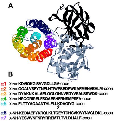
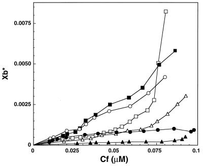
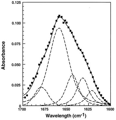
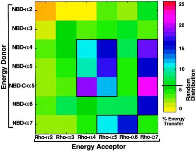
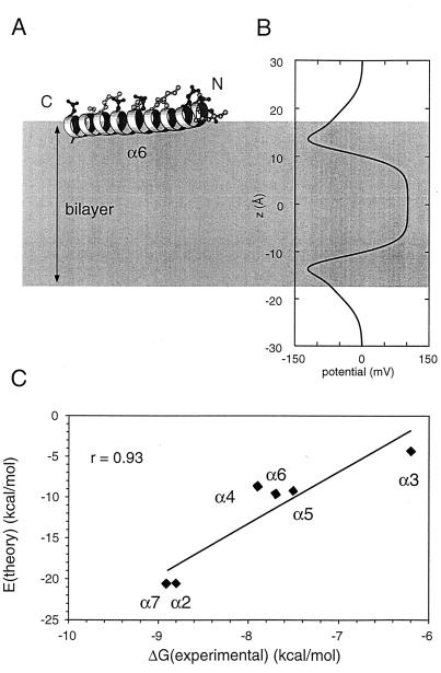
The aim of this study was to elucidate the mechanism of membrane insertion and the structural organization of pores formed by Bacillus thuringiensis delta-endotoxin. We determined the relative affinities for membranes of peptides corresponding to the seven helices that compose the toxin pore-forming domain, their modes of membrane interaction, their structures within membranes, and their orientations relative to the membrane normal. In addition, we used resonance energy transfer measurements of all possible combinatorial pairs of membrane-bound helices to map the network of interactions between helices in their membrane-bound state. The interaction of the helices with the bilayer membrane was also probed by a Monte Carlo simulation protocol to determine lowest-energy orientations. Our results are consistent with a situation in which helices alpha4 and alpha5 insert into the membrane as a helical hairpin in an antiparallel manner, while the other helices lie on the membrane surface like the ribs of an umbrella (the "umbrella model"). Our results also support the suggestion that alpha7 may serve as a binding sensor to initiate the structural rearrangement of the pore-forming domain.


Figures

Figure 1
Figure 1
Structure of B. thuringiensis δ-endotoxin. (A) Schematic ribbon representations of the CryIIIA toxin showing the domain organization as determined by ref. : domain I (colored), the pore-forming domain; domain II (gray), the receptor-binding domain; and domain III (black). The helices of the pore-forming domain are colored in a rainbow direction: α1, red; α2, orange; α3, yellow; α4, green; α5, cyan; α6, blue; and α7, purple. These illustrations were made by using the rasmol program. (B) Sequences of the pore-forming helices and their corresponding synthetic peptides. For unlabeled peptides, X = H and Z = H. For labeled peptides, X = NBD or Rho. Only for NBD-C-α5, X = acetyl and Z = NBD (14, 15).
Figure 2
Figure 2
Isotherms for binding of NBD-labeled α2–α7 peptides to phospholipid vesicles. The binding isotherms were derived from binding curves of NBD-labeled peptides titrated with phospholipid vesicles as described in ref. . The binding isotherms of α5 and α7 are from refs. and , respectively. ■, NBD-α2; ▴, NBD-α3; •, NBD-α4; □, NBD-α5; ▵, NBD-α6; and ○, NBD-α7.
Figure 3
Figure 3
Spectral deconvolution of the amide I band of α2 peptide incorporated into phospholipid membranes. The component peaks are the result of curve-fitting using a Voigt line-shape. The amide I frequencies characteristic of the various secondary structure elements are from ref. . The sum of the fitted components superimposes on the experimental amide I region spectrum. Filled squares, experimental Fourier-transform IR spectrum; dashed lines, the fitted components; solid line, the sum of the fitted components.
Figure 4
Figure 4
Theoretically and experimentally derived percentage of energy transfer versus bound-acceptor/lipid molar ratios. The amount of lipid-bound acceptor (Rho-peptides), Cb, at various acceptor concentrations was calculated from the binding isotherms. •, NBD-C-α5/Rho-α7; ▵, NBD-C-α5/Rho-α4; ▴, NBD-α5/Rho-α4; ■, NBD-C-α5/Rho-α5; □, NBD-C-α5/Rho-α6; ○, NBD-C-α5/Rho-α3; ▿, NBD-C-α5/Rho-α2; - - -, energy transfer expected for random distribution of the monomers (29), assuming R0 = 51.1 Å as determined for membrane-bound NBD-Rho pair (13). Results are shown as mean ± SE, n = 4, except for NBD-C-α5 ↔ Rho-α4 and NBD-α5 ↔ Rho-α4, in which n = 7 for each experimental point.
Figure 5
Figure 5
Fluorescence energy transfer between NBD-labeled peptides (donor) and Rho-labeled peptides (acceptors). The degree of energy transfer between acceptor and donor peptides of the 42 possible combinatorial acceptor–donor pairs is represented as a pseudocolor map. The energy transfer was calculated as in Fig. 4. Thick line marks the areas where the energy transfer was significantly higher than expected for random distribution of monomers.
Figure 6
Figure 6
MC simulations of peptide/bilayer interactions. (A) The α6 helix (drawn by using molscript). (B) Electrostatic potential profile, Φ(z), used in the EHEADGROUP term of the interaction energy function. The gray area indicates the extent (|z| < 17 Å) of the bilayer potential used in the simulations. (C) Minimum potential energy of interaction (EMIN) for each peptide plotted against its experimentally estimated free energy of interaction with a phospholipid bilayer. The best-fit line (r = 0.93) is shown superimposed on the data points.
Figure 7
Figure 7
Schematic presentation of a proposed model for the interaction of δ-endotoxin with phospholipid membranes. The helices are colored in a rainbow direction as in Fig. 1. The loop connecting α4 and α5 may be either in an intracellular localization or may interact with the inner leaflet of the membrane because of its hydrophobicity.

References

    1. Knowles B H. Adv Insect Physiol. 1994;24:275–308.
    1. Li J D, Carroll J, Ellar D J. Nature (London) 1991;353:815–821. - PubMed
    1. Lemmon M A, Engelman D M. Q Rev Biophys. 1994;27:157–218. - PubMed
    1. Van Rie J, McGaughey W H, Johnson D E, Barnett B D, Van Mellaert H. Science. 1990;247:72–74. - PubMed
    1. Ahmad W, Ellar D J. FEMS Microbiol Lett. 1990;68:97–104. - PubMed

Publication types

LinkOut - more resources