. 1998 Oct 13;95(21):12300-5.
doi: 10.1073/pnas.95.21.12300.

Protein thermostability above 100 degreesC: a key role for ionic interactions

Affiliations

Protein thermostability above 100 degreesC: a key role for ionic interactions

C Vetriani et al. Proc Natl Acad Sci U S A. .

Abstract

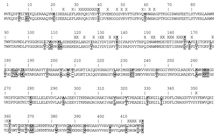
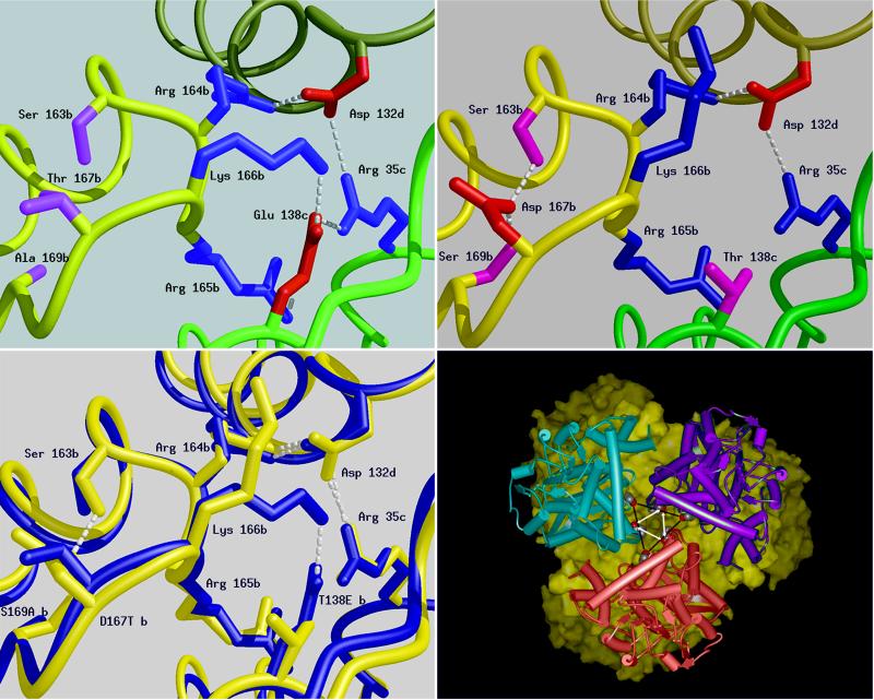
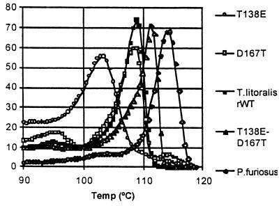
The discovery of hyperthermophilic microorganisms and the analysis of hyperthermostable enzymes has established the fact that multisubunit enzymes can survive for prolonged periods at temperatures above 100 degreesC. We have carried out homology-based modeling and direct structure comparison on the hexameric glutamate dehydrogenases from the hyperthermophiles Pyrococcus furiosus and Thermococcus litoralis whose optimal growth temperatures are 100 degreesC and 88 degreesC, respectively, to determine key stabilizing features. These enzymes, which are 87% homologous, differ 16-fold in thermal stability at 104 degreesC. We observed that an intersubunit ion-pair network was substantially reduced in the less stable enzyme from T. litoralis, and two residues were then altered to restore these interactions. The single mutations both had adverse effects on the thermostability of the protein. However, with both mutations in place, we observed a fourfold improvement of stability at 104 degreesC over the wild-type enzyme. The catalytic properties of the enzymes were unaffected by the mutations. These results suggest that extensive ion-pair networks may provide a general strategy for manipulating enzyme thermostability of multisubunit enzymes. However, this study emphasizes the importance of the exact local environment of a residue in determining its effects on stability.


Figures

Figure 1
Figure 1
Sequence alignment of archaeal GluDH from P. furiosus (upper) and T. litoralis (lower) with sequence differences shown in bold and underlined. Intersubunit contacts closer than 3.7 Å within the P. furiosus hexamer are indicated above the sequences (X). Contact residues that are variable (X) include S40T, T138E, D167T, D362Y, and K419H.
Figure 2
Figure 2
Schematic diagrams of a six residue-charged cluster in the structure of the GluDHs formed between residues from three adjacent subunits produced by the program midas (31). The main chain of each subunit is shown as a smooth ribbon with b, c, and d subunits colored yellow, green, and khaki, respectively. The positively charged side chains are shown in blue, the negatively charged ones in red, and hydrogen bonds shown as dashed lines. (Upper Left) P. furiosus GluDH: Glu-138, subunit c can be seen to form triple ion-pair interactions to Arg-35, subunit c; Arg-165, subunit b; and Lys-166, subunit b. (Upper Right) The same cluster in T. litoralis GluDH, highlighting the difference caused by the substitution in this enzyme of Glu-138 by Thr (shown in pink). The main consequence of this cluster is the reduction in the size of the charged network from six residues to three. The transposition of residues 138 and 167 leads to very little change in the position of neighboring residues with the notable exception of K166, which in T. litoralis is found extended toward the inter-trimer interface rather than between the subunits of the trimer. D167 (T. litoralis) is unshielded and its carboxyl groups are buried away from the lumen on the threefold axis. (Lower Left) A superimposition of the structures of the GluDHs from T. litoralis (blue) and P. furiosus (red). This diagram highlights the difference between the two structures in the region of the sequence change at position 138. Subtle displacement of the main chain occurs with the two backbone loops of the P. furiosus GluDH further apart at position 167. (Lower Right) View along the threefold axis of T. litoralis GluDH hexamer was generated by using msi WebLab. One trimer is depicted as a solid surface (yellow) and the other as three monomers (pink, blue, and purple). The mutation sites side chains are indicated by the vertices of the central triangles (T138 black bars, D167 white arrows). The corresponding sites on the surface of the other trimer are juxtaposed.
Figure 3
Figure 3
Relative heat capacity of GluDHs between 90°C and 130°C. The thermal stability of the double mutant T138E/D167T is elevated relative to T. litoralis wild-type and approaches that of the native P. furiosus GluDH. The single mutants T138E and D167T are relatively less stable, acutely so in the case of T138E, which has an extremely gradual transition.

References

    1. Adams M W. Annu Rev Microbiol. 1993;47:627–658. - PubMed
    1. Chan M K, Mukund S, Kletzin A, Adams M W, Rees D C. Science. 1995;267:1463–1469. - PubMed
    1. Anderson D E, Hurley J H, Nicholson H, Baase W A, Matthews B W. Protein Sci. 1993;2:1285–1290. - PMC - PubMed
    1. Hurley J H, Baase W A, Matthews B W. J Mol Biol. 1992;224:1143–1159. - PubMed
    1. Britton K L, Baker P J, Borges K M, Engel P C, Pasquo A, Rice D W, Robb F T, Scandurra R, Stillman T J, Yip K S P. Eur J Biochem. 1995;229:688–695. - PubMed

Publication types

Associated data

LinkOut - more resources