. 1998 Oct 13;95(21):12334-9.
doi: 10.1073/pnas.95.21.12334.

A DNA damage and stress inducible G protein-coupled receptor blocks cells in G2/M

Affiliations

A DNA damage and stress inducible G protein-coupled receptor blocks cells in G2/M

Z Weng et al. Proc Natl Acad Sci U S A. .

Abstract

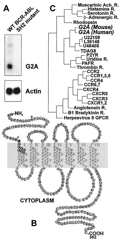
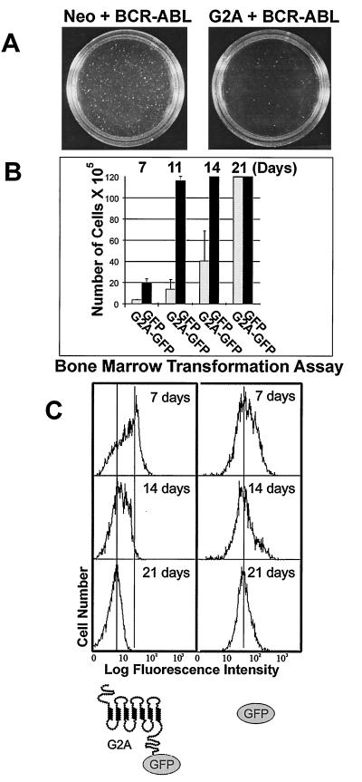
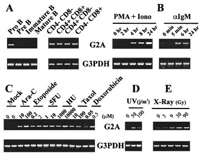
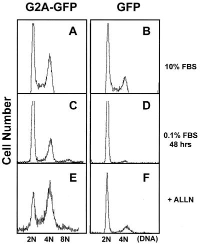
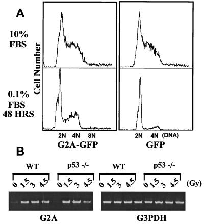
Cell cycle progression is monitored by highly coordinated checkpoint machinery, which is activated to induce cell cycle arrest until defects like DNA damage are corrected. We have isolated an anti-proliferative cell cycle regulator named G2A (for G2 accumulation), which is predominantly expressed in immature T and B lymphocyte progenitors and is a member of the seven membrane-spanning G protein-coupled receptor family. G2A overexpression attenuates the transformation potential of BCR-ABL and other oncogenes, and leads to accumulation of cells at G2/M independently of p53 and c-Abl. G2A can be induced in lymphocytes and to a lesser extent in nonlymphocyte cell lines or tissues by multiple stimuli including different classes of DNA-damaging agents and serves as a response to damage and cellular stimulation which functions to slow cell cycle progression.


Figures

Figure 1
Figure 1
G2A is a BCR-ABL-induced GPCR. (A) Induction of G2A by wt BCR-ABL but not by the SH2 mutant of BCR-ABL. Murine bone marrow cells were infected with retrovirus encoding the wt BCR-ABL or the SH2 mutant (R552L) expressed from the long terminal repeat promoter in the pSRαMSV vector (35, 36, 79). Transformed pre-B-lymphoid cells were harvested, and RNA was extracted for Northern blotting. The major G2A transcript is 3 kb in length. Actin was used as a total RNA-loading control. (B) Predicted transmembrane helices of the murine G2A. The potential glycosyslation site (∗) and the putative destruction box (arrow) are indicated. Sequence analysis was performed by using the blast program (Genetics Computer Group, Madison, WI). The transmembrane helices were predicted by using the sosui program available at http://expasy.hcuge.ch/www/tools.html. (C). G2A belongs to a subclass of transcriptionally regulated GPCRs. Phylogenetic analysis of G2A was made with the program pileup (Genetics Computer Group). Phylogenetic relationships were calculated with distances (Jukes–Cantor Distance algorithm) and the tree was made with growtree (UPGMA algorithm).
Figure 2
Figure 2
G2A antagonizes the oncogenic potential of the BCR-ABL oncogene. (A) Ectopic expression of G2A attenuates the proliferative signals of BCR-ABL in a fibroblast transformation assay. G2A with the neomycin resistance gene (neo) or Neo only expressing rat fibroblast (Rat-1) cell lines were generated by retroviral infection and G418 selection and were then super-infected with a retroviral stock expressing p185 BCR-ABL. The cells were plated in agar as described (48) and colonies were counted and photographed after 3 weeks. (B) G2A suppresses the induction of pre-B cells by BCR-ABL in bone marrow cells. Bone marrow transformation was performed as described (51) by using bone marrow cells from the tibias and femurs of BALB/c mice. Bone marrow was harvested and infected with retrovirus encoding the wt p185 BCR-ABL along with the G2A-GFP fusion protein (pMSCV G2A-GFP IRES p185 wt) or the GFP control (pMSCV GFP IRES p185 wt). The cells were plated in triplicate at a density of 5 × 10⁶ cells per 6-cm dish in RPMI medium 1640 containing 10% fetal bovine serum and 36-mercapto-ethanol (5 × 10⁻⁵ M, Sigma) and monitored for pre-B cell growth at the various time points indicated. Bone marrow cultures reached confluency around 1 × 10⁷ cells/per 3 ml culture. (C) Counter-selection against the G2A protein level in bone marrow cells transformed by BCR-ABL. The protein level of G2A-GFP or GFP was monitored by FACS in BCR-ABL-transformed bone marrow cells at time points indicated.
Figure 3
Figure 3
Multiple stimuli induce G2A. Mouse bone marrow B cells were isolated, stained with the B220, CD43, BP-1, IgM, and IgD antibodies, and FACS sorted into pro-B (B220+, CD43+), pre-B (B220+, BP1+, CD43−), immature B (B220+, IgM+, IgD−), and mature B cells (B220+, IgM+, IgD+). Similarly, mouse thymocytes were isolated from thymus, stained with CD4 and CD8 antibodies, and fractionated into CD4-CD8−, CD4+CD8+, CD4+CD8−, or CD4-CD8+ populations. RNA samples were extracted from 105 of cells of each fraction, and RT-PCR was then performed. A semi-quantitative RT-PCR method was used to measure the RNA levels of G2A (see Materials and Methods). Glyceraldehyde 3-phosphate dehydrogenase was used as a control to ensure that equal amounts of templates were used for RT-PCR. (B). Human Ramos B cells (54) were activated by either phorbol 12-myristate 13-acetate (PMA 2, ug/ml) plus ionomycin (20 ng/ml) or goat anti-IgM (final concentration of 10 μg/ml, Southern Biotechnology Associates). RNA samples were isolated before or after activation at time points indicated. RT-PCR was then performed by using primers specific for the human G2A cDNA sequence. (CE) Induction of G2A in B cells by different classes of DNA-damaging agents: Ramos cells were irradiated with increasing doses of x-ray, UV, or chemotherapy drugs and then were grown in medium for additional 16 hr before RNA samples were extracted for RT-PCR.
Figure 4
Figure 4
Ectopic expression of G2A accumulates cells at G2/M and blocks the progression of mitosis. Retroviruses expressing either a G2A-GFP fusion or a GFP control were used to infect NIH 3T3 cells. NIH 3T3 cells expressing G2A-GFP or GFP control were grown in 10% serum (A and B) or starved in media with 0.1% FBS for 48 hr (C and D). Cells were then harvested, and the DNA content was analyzed. Mouse fibroblasts were infected with retroviruses expressing G2A-GFP or GFP and then incubated with 50 μM of ALLN overnight in media with 10% FBS. The cells were harvested and analyzed for DNA content (E and F).
Figure 5
Figure 5
p53 is not required for the G2/M block by G2A or the induction of G2A. (A) Retroviruses expressing either G2A-GFP fusion, or a GFP control were used to infect p53−/− fibroblasts (61). Cells were cultured in the presence of 10% or 0.1% FBS for 48 hr and harvested for DNA content analysis by FACS. (B) Bone marrow cells were isolated from the wt and p53-knock-out mice and incubated with interleukin-7 and stem cell factor to stimulate the outgrowth of pre-B cells (63). After 3 days, the pre-B cells were irradiated with varying doses of x-ray followed by overnight incubation. RT-PCR analysis was then performed (see Materials and Methods).

References

    1. Hartwell L H, Weinert T A. Science. 1989;246:629–634. - PubMed
    1. Hartwell L. Cell. 1992;71:543–546. - PubMed
    1. Murray A W. Nature (London) 1992;359:599–604. - PubMed
    1. Hunter T. Cell. 1993;75:839–841. - PubMed
    1. Hartwell L H, Kastan M B. Science. 1994;266:1821–1828. - PubMed

Publication types

Associated data