. 1998 Oct 13;95(21):12352-7.
doi: 10.1073/pnas.95.21.12352.

Nuclear-encoded proteins target to the plastid in Toxoplasma gondii and Plasmodium falciparum

Affiliations

Nuclear-encoded proteins target to the plastid in Toxoplasma gondii and Plasmodium falciparum

R F Waller et al. Proc Natl Acad Sci U S A. .

Abstract

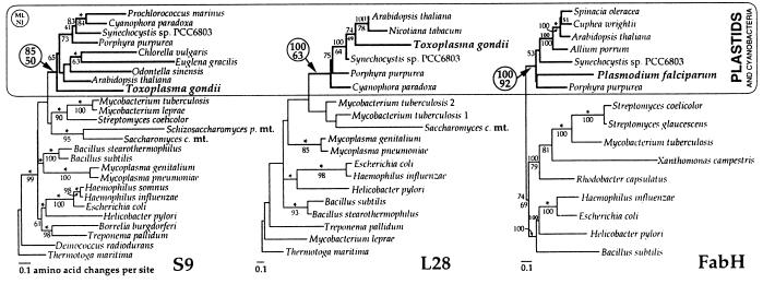
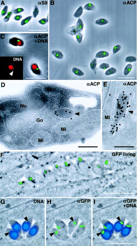
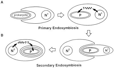
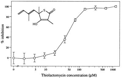
A vestigial, nonphotosynthetic plastid has been identified recently in protozoan parasites of the phylum Apicomplexa. The apicomplexan plastid, or "apicoplast," is indispensable, but the complete sequence of both the Plasmodium falciparum and Toxoplasma gondii apicoplast genomes has offered no clue as to what essential metabolic function(s) this organelle might perform in parasites. To investigate possible functions of the apicoplast, we sought to identify nuclear-encoded genes whose products are targeted to the apicoplast in Plasmodium and Toxoplasma. We describe here nuclear genes encoding ribosomal proteins S9 and L28 and the fatty acid biosynthetic enzymes acyl carrier protein (ACP), beta-ketoacyl-ACP synthase III (FabH), and beta-hydroxyacyl-ACP dehydratase (FabZ). These genes show high similarity to plastid homologues, and immunolocalization of S9 and ACP verifies that the proteins accumulate in the plastid. All the putatively apicoplast-targeted proteins bear N-terminal presequences consistent with plastid targeting, and the ACP presequence is shown to be sufficient to target a recombinant green fluorescent protein reporter to the apicoplast in transgenic T. gondii. Localization of ACP, and very probably FabH and FabZ, in the apicoplast implicates fatty acid biosynthesis as a likely function of the apicoplast. Moreover, inhibition of P. falciparum growth by thiolactomycin, an inhibitor of FabH, indicates a vital role for apicoplast fatty acid biosynthesis. Because the fatty acid biosynthesis genes identified here are of a plastid/bacterial type, and distinct from those of the equivalent pathway in animals, fatty acid biosynthesis is potentially an excellent target for therapeutics directed against malaria, toxoplasmosis, and other apicomplexan-mediated diseases.


Figures

Figure 1
Figure 1
Phylogenetic relationship of apicomplexan S9, L28, and FabH proteins to plastid and cyanobacterial homologs. Neighbor-joining (NJ) trees are shown with bootstrap confidence values >50% from both NJ (below the nodes) and protein maximum likelihood (ML) (above the nodes). Asterisks indicate constrained nodes in ML analyses. For all three data sets, the apicomplexan genes consistently group among genes from plastids and cyanobacteria (see circled bootstrap values) and distinct from mitochondria (mt data available only for S9 and L28) and other eubacterial groups.
Figure 2
Figure 2
Apicoplast localization of nuclear-encoded proteins. (A and B) Immunodetection of S9 and ACP (respectively) in intact T. gondii cells demonstrates that these proteins are restricted to a distinct region of the parasite similar to the location of the apicoplast. Some cells show two plastids, which are probably division stages. (C) Counterstaining with propidium iodide (red) confirms colocalization of ACP (green) with the extranuclear apicoplast DNA (Inset shows DNA staining only; arrowhead indicates apicoplast DNA). (D and E) Detection of ACP by immunogold labeling of ultra-thin sections shows strong labeling of the apicoplast (arrowhead). Nu, nucleus; Mi, mitochondrion; Go, Golgi apparatus. (F) The N-terminal domain of ACP (TgACPleader-GFP) is sufficient to target GFP to the apicoplast, and the recombinant protein can be visualized in living cells. (G–I) In fixed cells labeled with anti-GFP (green) and counterstained with DAPI (blue), TgACPleader-GFP can be seen to colocalize with the apicoplast DNA (arrowheads indicate apicoplasts in two cells). Color images were collected independently and overlaid on top of phase-contrast micrographs. (White scale bars = 10 μm, black scale bars = 200 nm).
Figure 3
Figure 3
Plastid origins and protein targeting. (A) Primary endosymbiosis describes the uptake of a prokaryote by a eukaryote. Plastids derived by primary endosymbiosis are generally surrounded by two membranes, and targeting of nuclear-encoded gene products to the endosymbiont is effected by an N-terminal transit peptide (T). (B) Secondary endosymbiotic plastid origin involves a heterotrophic eukaryote phagocytozing a photosynthetic eukaryote produced by primary endosymbiosis. The secondary endosymbiont’s cytoplasm and nucleus (N1) are typically lost or heavily reduced, and the resulting plastid is surrounded by four membranes, the outermost of which derives from the phagocytotic membrane. Sometimes one of the two outer membranes is lost at this point, resulting in a total of three. Targeting of nucleus-encoded (N2) gene products to secondary plastids requires a signal peptide (S) to mediate protein passage across the outer membrane(s) followed by a transit peptide (T) for import across the inner membrane pair.
Figure 4
Figure 4
Schematic of inferred proteins of T. gondii (Tg) genes rps9, rpl28, fabZ, and acpP and P. falciparum (Pf) genes acpP and fabH, showing N-terminal extensions resembling signal plus transit peptides (shaded and black boxes, respectively). Putative signal peptide cleavage sites were predicted by using signalp and psort (18); transit peptide/mature protein boundaries were imprecisely defined based on Western data (see Fig. 5) and sequence similarity with other mature proteins. Intron positions are indicated (triangle) (except for FabZ); “ψ” and “∗” indicate introns shared between T. gondii and P. falciparum homologs, suggesting shared ancestry.
Figure 5
Figure 5
Processing of apicoplast-targeted proteins in T. gondii. (A) Schematic of T. gondii S9 and ACP proteins, and GFP fusion proteins (ACPfull-GFP and ACPleader-GFP), showing protein band sizes before and after predicted processing (mature ACP carries a phosophopantetheine prosthetic group that adds 4 kDa to its expected gel mobility (45). (B) Western blot analysis of T. gondii S9 and ACP proteins (lanes i and ii, respectively). The observed antigens are in close agreement with the expected sizes for fully processed protein (lower bands) and full-length protein including the transit peptide but lacking the signal sequence (upper bands). (C) Western blot analysis of TgACPfull-GFP and TgACPleader-GFP proteins (lanes iii and iv). Observed molecular masses correspond to the predictions shown in (A) (the faint lower molecular mass band of ≈30 kDa in the ACPfull-GFP lane is not seen in untransfected parasites and probably represents a GFP degradation product). Note that similar amounts and relative levels of processed and unprocessed apicoplast proteins are observed for the native and recombinant proteins.
Figure 6
Figure 6
Log concentration-growth inhibition curves for the type II fatty acid biosynthesis inhibitor, thiolactomycin, tested against P. falciparum. The IC50 for both D10-strain and the multi-drug-resistant strain W2 mef (data not shown) P. falciparum was calculated as ≈50 μM. Vertical bars indicate standard deviations from six samples. (Inset) the structure of thiolactomycin.

References

    1. Wilson R J M, Denny P W, Preiser P R, Rangachari K, Roberts K, Roy A, Whyte A, Strath M, Moore D J, Moore P W, et al. J Mol Biol. 1996;261:155–172. - PubMed
    1. McFadden G I, Reith M, Munholland J, Lang-Unnasch N. Nature (London) 1996;381:482. - PubMed
    1. Köhler S, Delwiche C F, Denny P W, Tilney L G, Webster P, Wilson R J M, Palmer J D, Roos D S. Science. 1997;275:1485–1489. - PubMed
    1. Denny P, Preisser P, Williamson D, Wilson I. Protist. 1998;149:51–59. - PubMed
    1. Blunt D, Kjramtsov N, Upton S, Montelone B. Clin Diagn Lab Immunol. 1997;4:11–13. - PMC - PubMed

Publication types

LinkOut - more resources