. 1998 Oct 13;95(21):12370-5.
doi: 10.1073/pnas.95.21.12370.

Delayed embryonic lethality in mice lacking protein phosphatase 2A catalytic subunit Calpha

Affiliations

Delayed embryonic lethality in mice lacking protein phosphatase 2A catalytic subunit Calpha

J Götz et al. Proc Natl Acad Sci U S A. .

Abstract

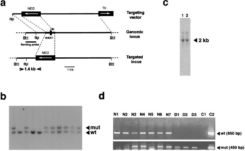
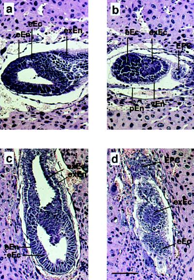
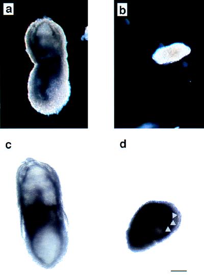
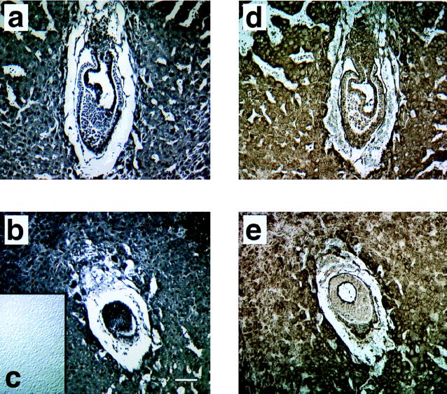
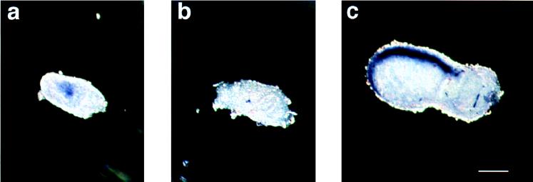
Protein phosphatase 2A (PP2A) is a multimeric enzyme, containing a catalytic subunit complexed with two regulatory subunits. The catalytic subunit PP2A C is encoded by two distinct and unlinked genes, termed Calpha and Cbeta. The specific function of these two catalytic subunits is unknown. To address the possible redundancy between PP2A and related phosphatases as well as between Calpha and Cbeta, the Calpha subunit gene was deleted by homologous recombination. Homozygous null mutant mice are embryonically lethal, demonstrating that the Calpha subunit gene is an essential gene. As PP2A exerts a range of cellular functions including cell cycle regulation and cell fate determination, we were surprised to find that these embryos develop normally until postimplantation, around embryonic day 5.5/6.0. While no Calpha protein is expressed, we find comparable expression levels of PP2A C at a time when the embryo is degenerating. Despite a 97% amino acid identity, Cbeta cannot completely compensate for the absence of Calpha. Degenerated embryos can be recovered even at embryonic day 13.5, indicating that although embryonic tissue is still capable of proliferating, normal differentiation is significantly impaired. While the primary germ layers ectoderm and endoderm are formed, mesoderm is not formed in degenerating embryos.


Figures

Figure 1
Figure 1
PP2A Cα gene targeting and genotyping. (a) Homologous recombination of the Cα genomic locus resulted in the replacement of 0.5 kb of the promoter, the entire exon I, and 0.3 kb of intron I sequences by the neomycin cassette. The neomycin cassette was flanked by 1 kb and 6 kb of genomic Cα sequences, respectively. The transcriptional orientation of the Cα gene is indicated by an arrow. BHI, BamHI; Bgl, BglII. The diagnostic primers WO-1 and P3 used to detect homologous recombination are indicated by arrowheads. (b) Southern blot analysis of viable offspring obtained from heterozygous matings. Genomic DNA was digested with BamHI (BHI) and probed with a flanking BamHI/BglII fragment (shown in a). Viable offspring were either heterozygous (+/−) or wild-type (+/+) as shown by the presence of a 9-kb wild-type and 10.1-kb mutant band, respectively. (c) Northern blot analysis of RNA obtained from wild-type (lane 1) and heterozygous (lane 2) brain, using a Cα-specific antisense oligonucleotide probe. (d) Genotyping of dissected E8.5 embryos obtained after heterozygous matings. All degenerated embryos (D1–D3) were genotypically −/− as shown by the presence of a 450-bp mutant PCR product and the absence of a 650-bp wild-type PCR product; all phenotypically normal embryos (N1–N7) were either +/+ or +/− as shown by the presence of a 650-bp wild-type PCR product. Two controls are shown for the two PCRs: For the wild-type PCR, targeting plasmid ZH13 was included as a negative control (C1) and wild-type tail lysate (C2) as a positive control. As controls for the presence of the mutant allele, wild-type (C1) and heterozygous (C2) tail lysates were included.
Figure 2
Figure 2
Morphology of embryos obtained from heterozygous crosses. Serial sagittal sections were made from embryonic day E6.5 (a and b) and E7.0 (c and d) deciduae (29). The genotype of these embryos was determined by PCR analysis of serial sections from which embryonic tissue was scraped off. Sections were stained with hematoxylin and eosin. Normal (a and c) and degenerated (b and d) embryos exhibit defined layers of cells of extra-embryonic (exEn) and embryonic (eEn) endoderm, and extra-embryonic (exEc) and embryonic (eEc) ectoderm. Degenerated embryos (b and d) are smaller and some of them are amorphous. Embryonic ectoderm seems to be more degenerated than extra-embryonic ectoderm (d). Parietal endoderm (pEn), visceral (vEn) endoderm, and the ectoplacental cone (EPC) are indicated. (Scale bar = 50 μm.)
Figure 3
Figure 3
Whole mount immunohistochemistry of dissected E7.5 embryos obtained from heterozygous matings. The genotype of dissected embryos was determined by PCR. (a and b) Dissected embryos were stained with antiserum 45 (17, 18), which is specific for Cα and does not cross-react with Cβ. In morphologically normal embryos (a) Cα is expressed almost uniformly, whereas morphologically abnormal (−/−) embryos (b) do not express Cα. (c and d) However, when an antiserum is employed that is directed against an epitope shared between Cα and Cβ, normal (c) and degenerated (d) embryos are stained equally well. Some of the degenerated embryos show a (proamniotic) cavity (d), the borders of which are indicated by arrowheads. (Scale bar = 50 μm.)
Figure 4
Figure 4
PP2A C (a–c) and Cβ (d and e) expression of embryos obtained from heterozygous crosses. Serial sagittal sections were made from E6.5 deciduae (29). The genotype of these embryos was determined by PCR analysis of serial sections from which embryonic tissue was scraped off. (a and b) Sections were stained with antiserum V598A, which is directed against an epitope common to Cα and Cβ. The degenerated embryo (b) exhibits the same staining intensity for C as the normal embryo (a). (Inset c) A control is included where the primary antiserum has been omitted. (d and e) Sections were stained with antiserum 15, which has been raised with a Cβ-specific peptide. Normal (d) and degenerated (e) embryos exhibit comparable staining intensities. (Scale bar = 50 μm.)
Figure 5
Figure 5
RNA whole mount in situ hybridization of embryos analyzed for the expression of mesodermal markers (23) by using digoxigenin-labeled antisense probes for goosecoid (data not shown) and Brachyury. (a–c) Embryos were dissected at E6.5 (a) and E7.0 (b and c). Transcripts (purple staining) are present in morphologically normal embryos (a and c) but are undetectable in degenerated embryos (b). (Scale bar = 50 μm.)

References

    1. Sontag E, Nunbhakdi-Craig V, Bloom G S, Mumby M C. J Cell Biol. 1995;128:1131–1144. - PMC - PubMed
    1. Snaith H A, Armstrong C G, Guo Y, Kaiser K, Cohen P T W. J Cell Sci. 1996;109:3001–3012. - PubMed
    1. Cohen P. Annu Rev Biochem. 1989;58:453–508. - PubMed
    1. Shenolikar S, Nairn A C. In: Advances in Second Messengers and Phosphoprotein Research. Greengard P, Robinson G A, editors. Vol. 25. New York: Raven; 1991. pp. 1–121. - PubMed
    1. Khew-Goodall Y, Hemmings B. FEBS Lett. 1988;238:265–268. - PubMed

Publication types