. 1998 Oct 13;95(21):12381-5.
doi: 10.1073/pnas.95.21.12381.

Natural genetic exchange between Haemophilus and Neisseria: intergeneric transfer of chromosomal genes between major human pathogens

Affiliations

Natural genetic exchange between Haemophilus and Neisseria: intergeneric transfer of chromosomal genes between major human pathogens

J S Kroll et al. Proc Natl Acad Sci U S A. .

Abstract

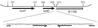
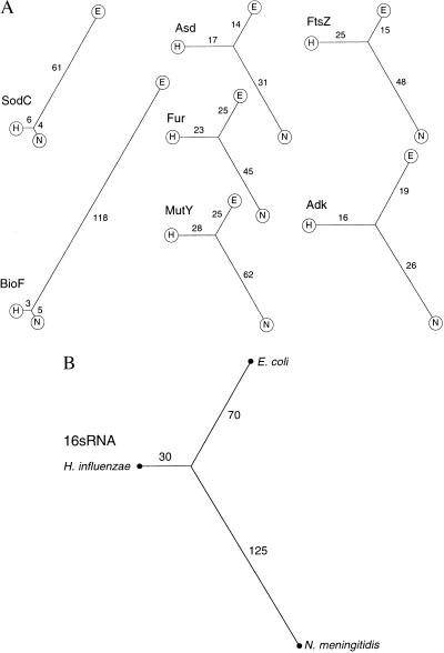
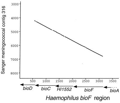
Members of the bacterial families Haemophilus and Neisseria, important human pathogens that commonly colonize the nasopharynx, are naturally competent for DNA uptake from their environment. In each genus this process is discriminant in favor of its own and against foreign DNA through sequence specificity of DNA receptors. The Haemophilus DNA uptake apparatus binds a 29-bp oligonucleotide domain containing a highly conserved 9-bp core sequence, whereas the neisserial apparatus binds a 10-bp motif. Each motif ("uptake sequence", US) is highly over-represented in the chromosome of the corresponding genus, particularly concentrated with core sequences in inverted pairs forming gene terminators. Two Haemophilus core USs were unexpectedly found forming the terminator of sodC in Neisseria meningitidis (meningococcus), and sequence analysis strongly suggests that this virulence gene, located next to IS1106, arose through horizontal transfer from Haemophilus. By using USs as search strings in a computer-based analysis of genome sequence, it was established that while USs of the "wrong" genus do not occur commonly in Neisseria or Haemophilus, where they do they are highly likely to flag domains of chromosomal DNA that have been transferred from Haemophilus. Three independent domains of Haemophilus-like DNA were found in the meningococcal chromosome, associated respectively with the virulence gene sodC, the bio gene cluster, and an unidentified orf. This report identifies intergenerically transferred DNA and its source in bacteria, and further identifies transformation with heterologous chromosomal DNA as a way of establishing potentially important chromosomal mosaicism in these pathogenic bacteria.


Figures

Figure 1
Figure 1
Shown is the 3,572-bp ClaI DNA insert of pJSK205. mutY and sodC are indicated by arrows, and the hatched rectangle indicates the region with homology to IS1106. Coordinates mark start/stop codons and the beginning of IS1106 homology, and correspond to those in GenBank accession no. AJ001313. The expansion below shows the sequence in intergenic region 3′ to sodC, with the inverted repeat highlighted. The sodC TAA stop codon and the first three bases of the region homologous to IS1106 are underlined.
Figure 2
Figure 2
Shown is the sequence of both strands of domain of pJSK205 between coordinates 2,270 and 2,301 (the region of the inverted repeat 3′ to sodC). Consensus HmUS is aligned with each strand [a, adenine in >50% HmUSs; n, any nucleotide; R, purine (A/G); W, weak (A/T)]. Matches are indicated by ∗.
Figure 3
Figure 3
(A) Phylograms based on protein sequence comparisons, computed with the darwin program phylotree of Gonnet et al. (32) accessed by e-mail (cbrg@inf.ethz.ch). In each case H represents H. influenzae (Hi), N = N. meningitidis (Nm), and E = E. coli (Ec). In the unrooted dendrograms, branch path length joining two peptide sequences (indicated) is in proportion to their degree of homology, calculated as the PAM (percentage accepted mutations) distance separating two sequences. Proteins (bacterial species, GenBank accession nos.) are represented as follows: Adk (Nm, L36469; Hi, X57315; Ec, X03038); Asd (Nm, Z14063; Hi, U32747; Ec, V00262); FtsZ (Nm, U43329; Hi, U32794; Ec, AE000119); Fur (Nm, L19777; Hi, U32704; Ec, X02589); MutY (Nm, this work; Hi, U32760; Ec, X52391); SodC (Nm, AJ001313; Hi, M84012; Ec, X97766); and BioF (Nm, Sanger sequence; Hi, U32830; Ec, AE000180). (B) Radial phylogram based on 16S RNA sequences, constructed from output of the computer program subtree of Maidak et al. (34) accessed at http://rdp.life.uiuc.edu. Branch lengths are in arbitrary units.
Figure 4
Figure 4
Graphical comparison of DNA sequences at the bio locus of H. influenzae (x-axis) and putative bio locus of N. meningitidis from Sanger contig 316 (y-axis). Points plotted represent minimum 80% sequence identity over a 30-nt moving window. Positions of known genes shown in relation to the Haemophilus sequence.
Figure 5
Figure 5
DNA sequence alignment of H. influenzae/N. meningitidis bioF with E. coli bioF in the region of the HmUS (underlined; arrow indicates consensus sequence on noncoding strand). Coding strands and their translations are shown: Haemophilus/Neisseria (identical to each other) above, E. coli below. Separation of the sequences represents divergence where Haemophilus/Neisseria contains the HmUS.

References

    1. Kilian M. J Gen Microbiol. 1976;93:9–62. - PubMed
    1. Danner D B, Deich R A, Sisco K L, Smith H O. Gene. 1980;11:311–318. - PubMed
    1. Goodgal S H, Mitchell M. J Bacteriol. 1984;157:785–788. - PMC - PubMed
    1. Smith H O, Tomb J F, Dougherty B A, Fleischmann R D, Venter J C. Science. 1995;269:538–540. - PubMed
    1. Fleischmann R D, Adams M D, White O, Clayton R A, Kirkness E F, Kerlavage A R, Bult C J, Tomb J F, Dougherty B A, Merrick J M, et al. Science. 1995;269:496–512. - PubMed

Publication types

Associated data

LinkOut - more resources