. 1998 Oct 13;95(21):12424-31.
doi: 10.1073/pnas.95.21.12424.

Apoptosis regulator bcl-w is essential for spermatogenesis but appears otherwise redundant

Affiliations

Apoptosis regulator bcl-w is essential for spermatogenesis but appears otherwise redundant

C G Print et al. Proc Natl Acad Sci U S A. .

Abstract

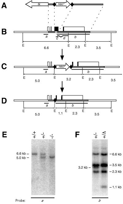
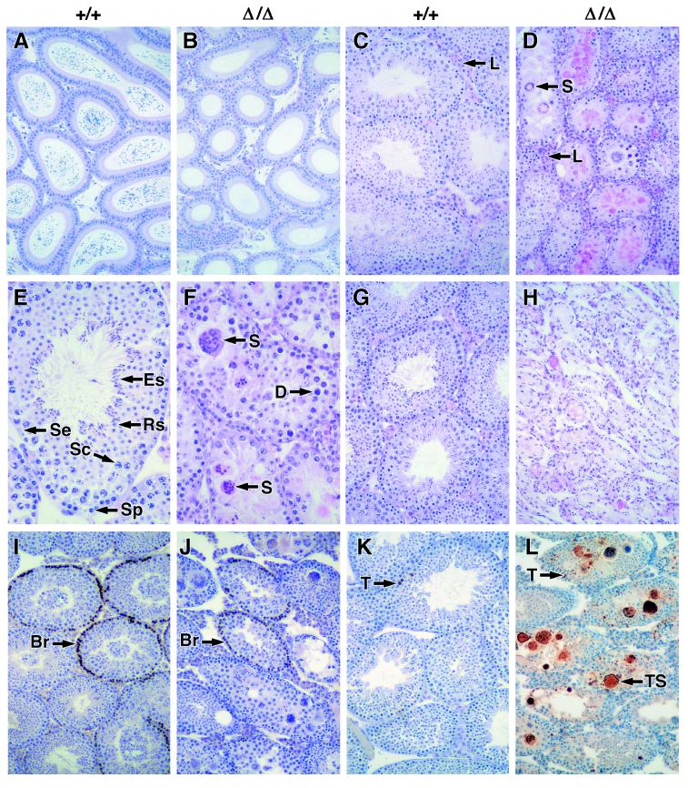
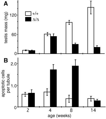
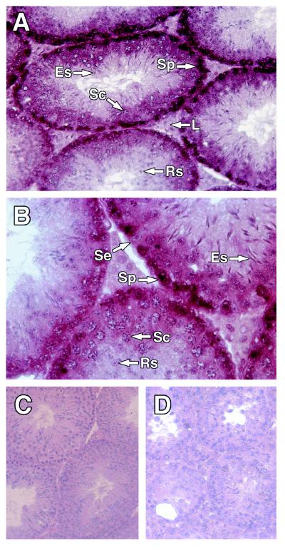
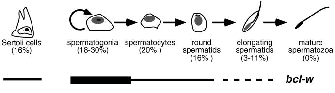
Proteins of the Bcl-2 family are important regulators of apoptosis in many tissues of the embryo and adult. The recently isolated bcl-w gene encodes a pro-survival member of the Bcl-2 family, which is widely expressed. To explore its physiological role, we have inactivated the bcl-w gene in the mouse by homologous recombination. Mice that lack Bcl-w were viable, healthy, and normal in appearance. Most tissues exhibited typical histology, and hematopoiesis was unaffected, presumably due to redundant function with other pro-survival family members. Although female reproductive function was normal, the males were infertile. The testes developed normally, and the initial, prepubertal wave of spermatogenesis was largely unaffected. The seminiferous tubules of adult males, however, were disorganized, contained numerous apoptotic cells, and produced no mature sperm. Both Sertoli cells and germ cells of all types were reduced in number, the most mature germ cells being the most severely depleted. The bcl-w-/- mouse provides a unique model of failed spermatogenesis in the adult that may be relevant to some cases of human male sterility.


Figures

Figure 1
Figure 1
Disruption of the bcl-w gene. (A) The targeting vector pbcl-wlox neor tk. Shaded bars represent regions derived from the bcl-w gene; tk, a thymidine kinase expression cassette; neor, a PGK- neor expression cassette; and diamonds, loxP sequences. (B) The wt bcl-w locus. Boxes represent exons (solid, coding region; open, untranslated region). E, EcoRI sites; sizes of EcoRI fragments are in kb. The bcl-w genomic DNA probes used for Southern blot analyses are labeled a and b, whereas the bcl-w cDNA sequences used as riboprobes are indicated by c and d. (C) Homologous recombination replaces the first 413 bp of the bcl-w coding region with a PGK-neor expression cassette bounded by loxP sites. (D) Cre-mediated recombination deletes the PGK-neor sequence, leaving only 127 bp of exogenous sequence, including a single loxP site. (E) Southern blot of genomic DNA from wt (+/+), heterozygous (+/−), and homozygous mutant (−/−) bcl-w mice (line 228), hybridized with bcl-w cDNA probe a. (F) Southern blot of genomic DNA from heterozygous mice (line 228) before (+/−) and after (+/Δ) the action of Cre recombinase, hybridized with bcl-w probe b.
Figure 2
Figure 2
Expression of the bcl-w gene. (A) Northern blot of total RNA (10 μg) extracted from the testes of 4-wk-old wt (+/+) and bcl-wΔ/Δ mice (Δ/Δ), hybridized to a probe containing the first 1.2 kb of the bcl-w cDNA (Upper); glyceraldehyde phosphate dehydrogenase mRNA served as a control (gapdh, Lower). (B) Western blot analysis of protein lysates from the brain, testis, and pancreas of wt and bcl-wΔ/Δ mice, using a polyclonal anti-Bcl-w antibody. The 21-kDa Bcl-w protein is indicated. (C) Western blots of protein lysates from testis cell lines, with the same antibody. GC-1 is a germ cell line derived from type B spermatogonia, TM4 a Sertoli cell line, and TM3 a Leydig cell line; all were obtained from the American Type Culture Collection.
Figure 3
Figure 3
Failed spermatogenesis in mice lacking Bcl-w. Comparison of wt (+/+) and bcl-wΔ/Δ (Δ/Δ) tissues. (A and B) Epididymides of 6-wk-old mice stained with hematoxylin/eosin (85-fold magnification). (C−F) Testis from 8-wk-old mice stained with hematoxylin/eosin (C and D, 85-fold; E and F, 340-fold). S, symplasts; D, degenerating nuclei; L, Leydig cells; Se, Sertoli cells; Sp, spermatogonia; Sc, spermatocytes; Rs, round spermatids; and Es, elongating spermatids. (G and H) Testis from 52-wk-old mice stained with hematoxylin/eosin (85-fold). (I and J) BrdUrd-labeled testis from 6-wk-old mice immunostained with anti-BrdUrd antibody and then counterstained with hematoxylin (85-fold); Br indicates spermatogonia that have incorporated BrdUrd. (K and L) Testis from 6-wk-old mice labeled by TUNEL and counterstained with hematoxylin (85-fold). T, TUNEL-labeled apoptotic nuclei; TS, TUNEL-labeled symplasts.
Figure 4
Figure 4
Reduced numbers of various cell types within the seminiferous tubules of bcl-wΔ/Δ mice. Frequencies of the indicated cell types was determined by the optical disector method for seven 6-wk-old wt mice and eight 6-wk-old bcl-wΔ/Δ mice. The percentage of the wt cell numbers remaining in the testes of bcl-wΔ/Δ mice is indicated. Error bars denote two SEM.
Figure 5
Figure 5
Degeneration of testis in bcl-wΔ/Δ mice. (A) Mean mass of testes (three mice per group). (B) TUNEL-labeled nuclei per tubule, counted at 2, 4, 8, and 14 wk (three mice per group). Error bars denote two SEM.
Figure 6
Figure 6
bcl-w expression in testis. (A−D) Paraffin-embedded sections of testis from 14-wk-old mice hybridized to bcl-w riboprobes and counterstained with hematoxylin. (A and B) wt testis hybridized with antisense bcl-w riboprobe c1 (A, 230-fold magnification; B, 460-fold). (C) wt testis hybridized to sense bcl-w riboprobe c2 (165-fold). (D) Testis from a bcl-wΔ/Δ mouse, hybridized to antisense bcl-w riboprobe c1 (165-fold).
Figure 7
Figure 7
Consequences of Bcl-w loss in the testis. The percentages of the Sertoli cells and the different types of germ cells remaining in bcl-wΔ/Δ mice are indicated. The expression pattern of the gene is indicated schematically; the broken line indicates that the extent of expression in late stages of germ cell development remains to be clarified (see text).

References

    1. Strasser A, Huang D C S, Vaux D L. Biochim Biophys Acta. 1997;1333:F151–F178. - PubMed
    1. Adams, J. M. & Cory, S. (1998) Science, in press.
    1. Chao D T, Korsmeyer S J. Annu Rev Immunol. 1998;16:395–419. - PubMed
    1. Oltvai Z N, Milliman C L, Korsmeyer S J. Cell. 1993;74:609–619. - PubMed
    1. Pan G H, O’Rourke K, Dixit V M. J Biol Chem. 1998;273:5841–5845. - PubMed

Publication types

LinkOut - more resources