. 1998 Oct 13;95(21):12456-61.
doi: 10.1073/pnas.95.21.12456.

Riboregulation in Escherichia coli: DsrA RNA acts by RNA:RNA interactions at multiple loci

Affiliations

Riboregulation in Escherichia coli: DsrA RNA acts by RNA:RNA interactions at multiple loci

R A Lease et al. Proc Natl Acad Sci U S A. .

Abstract

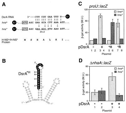
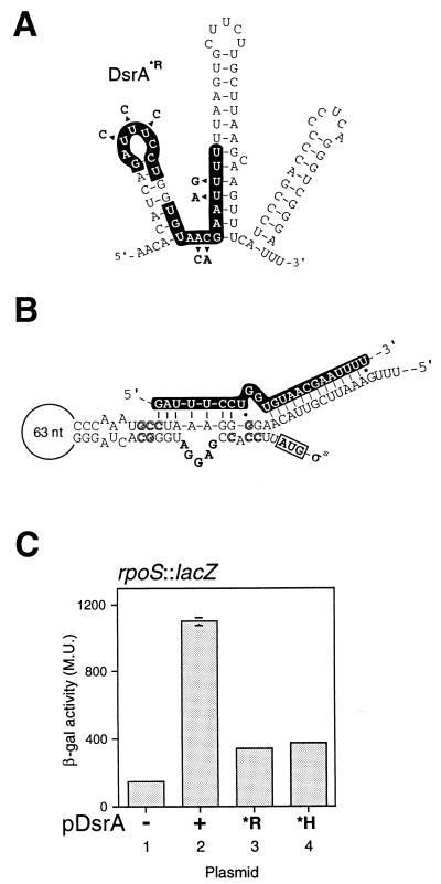
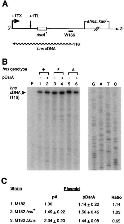
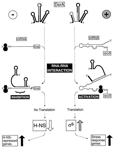
DsrA is an 87-nt untranslated RNA that regulates both the global transcriptional silencer and nucleoid protein H-NS and the stationary phase and stress response sigma factor RpoS (sigmas). We demonstrate that DsrA acts via specific RNA:RNA base pairing interactions at the hns locus to antagonize H-NS translation. We also give evidence that supports a role for RNA:RNA interactions at the rpoS locus to enhance RpoS translation. Negative regulation of hns by DsrA is achieved by the RNA:RNA interaction blocking translation of hns RNA. In contrast, results suggest that positive regulation of rpoS by DsrA occurs by formation of an RNA structure that activates a cis-acting translational operator. Sequences within DsrA complementary to three additional genes, argR, ilvIH, and rbsD, suggest that DsrA is a riboregulator of gene expression that acts coordinately via RNA:RNA interactions at multiple loci.


Figures

Figure 1
Figure 1
DsrA contains regions of complementarity to different genes in E. coli. Computer searches revealed complementarity to DsrA at or near the 5′ ends of the coding sequences of five E. coli genes. The DsrA nucleotide sequences complementary to indicated genes are highlighted (white letters on a black background) on the predicted structure of DsrA (13). Open arrowheads indicate potential G:U base pairs between DsrA and target sequences. A prime (′) after the gene name designates complementarity to the gene. (A) Complementarity to hns and rpoS demonstrated in this work. Two possible complementary schemes are shown for hns. (B) Putative complementarity to argR, ilvIH, and rbsD.
Figure 2
Figure 2
DsrA acts via RNA:RNA interactions to repress H-NS. (A) Silent mutagenesis of hns to disrupt complementarity to DsrA. Five nucleotides of wild-type hns were modified by site-directed mutagenesis (arrows) to disrupt the DsrA:hns RNA base pairing (vertical lines). The mutations, which are silent in terms of the H-NS protein sequence (below), create a StuI restriction site (underlined) in the altered hns* allele. The initiator methionine codon is indicated (boxed). (B) DsrA mutations that prevent base pairing with hns but allow base pairing with hns*. Mutated nucleotides are shown next to the DsrA structure (arrowheads). (C and D) Assay of H-NS function in strains with and without hns RNA:DsrA complementarity. DsrA-induced repression of H-NS transcription silencing activity was assayed in strain M182 by using a proUlacZ (C) or ΔnhaAlacZ (D) reporter gene. β-galactosidase assays were performed as described (25), except that assays on ΔnhaAlacZ used a buffered growth medium (37). All assays were performed in duplicate; values given are the average of three independent experiments. Light columns, hns+ strains; dark columns, hns* strains. Plasmids used: −, pA vector; +, pDsrA; *H, pDsrA*H as in B; *R, pDsrA*R as in Fig. 3A.
Figure 3
Figure 3
DsrA activates rpoS translation: an RNA:RNA interaction model. (A) DsrA*R mutant RNA. Mutations are indicated by arrowheads. (B) Proposed structure of the rpoS mRNA translational operator. Bold letters in the bulge loop indicate the Shine–Dalgarno sequence; boxed AUG indicates the initiator methionine codon; outlined letters indicate mutations that confirm secondary structure and enable Hfq-independent translation (adapted from ref. by using additional sequence of E. coli rpoS from ref. 48). The sequence of DsrA is given in white letters on a black background, indicating the complementarity to rpoS mRNA deduced in this work (Fig. 1A). Vertical lines are base pairs, dots are G:U base pairs. (C) Effect of altered DsrA on translation of rpoS mRNA. The reporter gene is a translational fusion between RpoS and LacZ in strain RO90. The RNA produced by the reporter fusion contains the region of DsrA complementarity. All values shown are the average of three independent experiments.
Figure 4
Figure 4
Levels of hns RNA during DsrA overexpression. (A) Diagram of the hns transcript and primer extension product. The hns primer W166 (20) hybridizes upstream of the deleted region in Δhns transcripts, enabling quantitation of RNA. +1 TX, transcriptional start; +1 TL, translational start. Size of primer extension product is given in nucleotides beside wavy line, representing cDNA. (B) Levels of hns RNA. RNA harvested from mid-log phase cells grown at 30°C was quantified by primer extension (29). Lanes and markers: P, no RNA; 1, M182hns+/pA; 2, M182hns+/pDsrA; 3, M182hns*/pA; 4, M182hns*/pDsrA; 5, M182Δhns/pA; 6, M182Δhns/pDsrA. The sequence of pTZ19U was used as a molecular weight marker (lanes G, A, T, and C). (C) Quantitation of hns cDNA primer-extension product. Values are normalized to M182hns+/pA. The average of three independent experiments, each performed in duplicate, is shown; error is reported as SD. All six data sets were subjected to variance analysis.
Figure 5
Figure 5
DsrA decreases H-NS and increases StpA levels in vivo. (A) Western blot of purified proteins and extracts fractionated by SDS/PAGE. The proteins were transferred to membranes and were probed with polyclonal antisera raised against H-NS. Lanes: 1 and 2, purified StpA and H-NS proteins, respectively; 3–7, crude lysates from M182/pA (lane 3), M182/pDsrA (lane 4), M182ΔstpA (lane 5), M182Δhns (lane 6), and M182Δstphns (lane 7). (B) H-NS and StpA proteins were separated by nonequilibrium two-dimensional isoelectric focusing and were visualized by Western blot analysis with mixed H-NS- and StpA-specific polyclonal antisera. The pH gradient is from ≈10 (left) to 5 (right). Cell extracts are from M182/pA, M182/pDsrA, and M182Δhns. (C) A separate experiment depicts H-NS separated by equilibrium two-dimensional isoelectric focusing.
Figure 6
Figure 6
Models for DsrA riboregulation. At the top, DsrA forms RNA:RNA interactions with target transcripts. On the left (−) is a model for translational repression of hns. On the right (+) is a model for translational activation of rpoS. Black circles represent ribosomes.

Similar articles

Cited by

References

    1. Rastinejad F, Conboy M J, Rando T A, Blau H M. Cell. 1993;75:1107–1117. - PubMed
    1. Simons R W, Kleckner N. Annu Rev Genet. 1988;22:567–600. - PubMed
    1. Delihas N. Mol Microbiol. 1995;15:411–414. - PubMed
    1. Henkin T M. Annu Rev Genet. 1996;30:35–57. - PubMed
    1. Moss E G, Lee R C, Ambros V. Cell. 1997;88:637–646. - PubMed

Publication types

LinkOut - more resources