. 1998 Oct 13;95(21):12492-7.
doi: 10.1073/pnas.95.21.12492.

Major histocompatibility complex (MHC) class I KbDb -/- deficient mice possess functional CD8+ T cells and natural killer cells

Affiliations

Major histocompatibility complex (MHC) class I KbDb -/- deficient mice possess functional CD8+ T cells and natural killer cells

Y Vugmeyster et al. Proc Natl Acad Sci U S A. .

Abstract

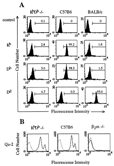
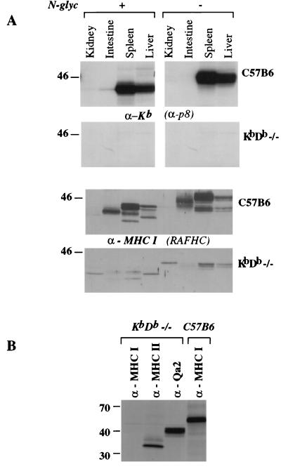
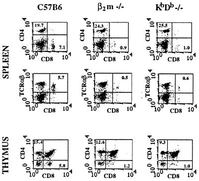
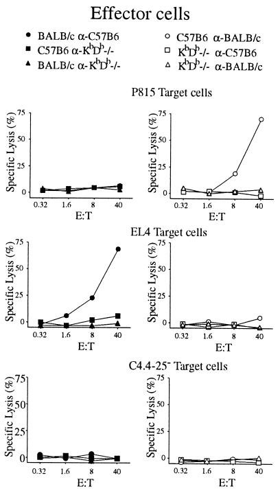
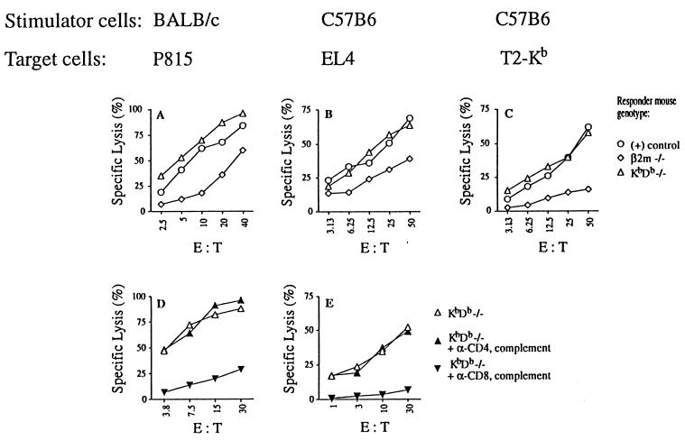
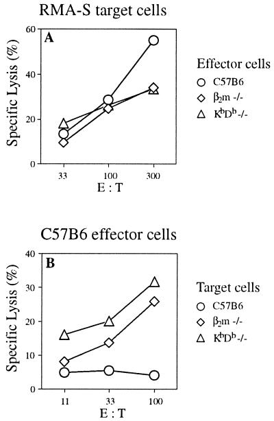
We obtained mice deficient for major histocompatibility complex (MHC) molecules encoded by the H-2K and H-2D genes. H-2 KbDb -/- mice express no detectable classical MHC class I-region associated (Ia) heavy chains, although beta2-microglobulin and the nonclassical class Ib proteins examined are expressed normally. KbDb -/- mice have greatly reduced numbers of mature CD8+ T cells, indicating that selection of the vast majority (>90%) of CD8+ T cells cannot be compensated for by beta2-microglobulin-associated molecules other than classical H-2K and D locus products. In accord with the greatly reduced number of CD8+ T cells, spleen cells from KbDb -/- mice do not generate cytotoxic responses in primary mixed-lymphocyte cultures against MHC-disparate (allogeneic) cells. However, in vivo priming of KbDb -/- mice with allogeneic cells resulted in strong CD8+ MHC class Ia-specific allogeneic responses. Thus, a minor population of functionally competent peripheral CD8+ T cells capable of strong cytotoxic activity arises in the complete absence of classical MHC class Ia molecules. KbDb -/- animals also have natural killer cells that retain their cytotoxic potential.


Figures

Figure 1
Figure 1
Cell-surface expression of MHC class Ia and class Ib molecules in KbDb −/− mice. (A) Blood lymphocytes from KbDb −/−, C57BL/6, or BALB/c mice were analyzed by direct immunofluorescence with α-Kb-FITC, α-Db-FITC, or α-Dd-FITC antibodies, as indicated. Numbers correspond to percentage of cells in the indicated regions. (B) Splenocytes from KbDb −/−, C57BL/6, or β2m −/− mice were analyzed by indirect immunofluorescence with biotinylated α-Qa-2 antibody and streptavidin-PE (solid line) or, as a control, with streptavidin-PE only (dashed line).
Figure 2
Figure 2
Expression of MHC-encoded molecules in KbDb −/− mice. (A) Whole-cell lysates from the indicated tissues were homogenized and adjusted for protein concentration (100 μg per lane). As a control, half of the samples were treated with N-glycosidase (Left). Lysates were analyzed by immunoblots with α-Kb (α-p8, Upper) or α-MHC class I free heavy chain (RAFHC, Lower) antibodies. (B) Spleen-cell Con A blasts were labeled biosynthetically for 45 min and chased for 2 h. Cell lysates were made, and H-2-encoded proteins were immunoprecipitated with α-Kb (α-p8), α-MHC class I free heavy chain (RAFHC), α-Qa-2, or α-MHC class II (N22) antibodies.
Figure 3
Figure 3
Reduction in CD8+TCRαβ T cell numbers in KbDb −/− mice. FACS analysis was performed on C57BL/6, β2m −/−, or KbDb −/− splenocytes (Top and Middle) or thymocytes (Bottom) with α-CD8-FITC, α-CD4-PE, and α-TCRαβ-PE antibodies, as indicated. Numbers correspond to percentage of cells in the indicated regions.
Figure 4
Figure 4
Absence of primary cytotoxic response in MLC by spleen cells from unprimed KbDb −/− mice. Effector cells were obtained by the standard 7-day MLC with spleen cells from C57BL/6, BALB/c, or KbDb −/− used as stimulators and responders (as indicated). CTL-mediated cytotoxicity was measured in a standard 4-h 51Cr release assay with H-2d P815 (Top), H-2b EL4 (Middle), or β2m −/− C4.4–25 (Bottom) targets. E:T, effector:target ratio.
Figure 5
Figure 5
Cytotoxic activity of CD8+ T cells from KbDb −/− mice after in vivo priming. C57BL/6, BALB/c, β2m −/−, or KbDb −/− mice were injected twice in vivo with either BALB/c (A and D) or C57BL/6 (B, C, and E) splenocytes. Effector cells were obtained by a standard 6-day MLC with the indicated splenocytes used as stimulators. KbDb −/− effector cells (anti-BALB/c in D and anti-C57BL/6 in E) were depleted of CD8+ or CD4+ T cells with α-CD8 or α-CD4 antibodies and complement (D and E). CTL-mediated cytotoxicity was measured in a standard 4-h 51Cr release assay using H-2d P815 (A and D), H-2b EL4 (B and E), or T2-Kb (C) target cells. The controls for A were C57BL/6 anti-BALB/c effectors, and the controls for B and C were BALB/c anti-C57BL/6 effectors (○).
Figure 6
Figure 6
KbDb −/− mice as donors of effector or target cells in NK-mediated cytotoxicity in vitro. (A) C57BL/6, β2m −/−, and KbDb −/− mice were injected with the interferon inducer tilorone, and splenocytes from these mice were used as effector cells 1 day after injection. NK-mediated cytotoxicity was measured with 4-h 51Cr release assay on RMA-S target cells. (B) C57BL/6 lymphokine-activated killer cells were obtained by 5-day culture of erythrocyte-depleted splenocytes. NK-mediated cytotoxicity was measured with 4-h 51Cr release assay on C57BL/6, β2m −/−, and KbDb −/− Con A blast target cells.

Similar articles

Cited by

References

    1. Bijlmakers M J, Neefjes J J, Wojcik-Jacobs E H, Ploegh H L. Eur J Immunol. 1993;23:1305–1313. - PubMed
    1. Jameson S C, Hogquist K A, Bevan M J. Annu Rev Immunol. 1995;13:93–126. - PubMed
    1. Ljunggren H G, Karre K. Immunol Today. 1990;11:237–244. - PubMed
    1. Tanchot C, Lemonnier F A, Perarnau B, Freitas A A, Rocha B. Science. 1997;276:2057–2062. - PubMed
    1. Markiewicz M A, Girao C, Opferman J T, Sun J, Hu Q, Agulnik A A, Bishop C E, Thompson C B, Ashton-Rickardt P G. Proc Natl Acad Sci USA. 1998;95:3065–3070. - PMC - PubMed

Publication types