. 1998 Oct 13;95(21):12580-5.
doi: 10.1073/pnas.95.21.12580.

Doxycycline control of prion protein transgene expression modulates prion disease in mice

Affiliations

Doxycycline control of prion protein transgene expression modulates prion disease in mice

P Tremblay et al. Proc Natl Acad Sci U S A. .

Abstract

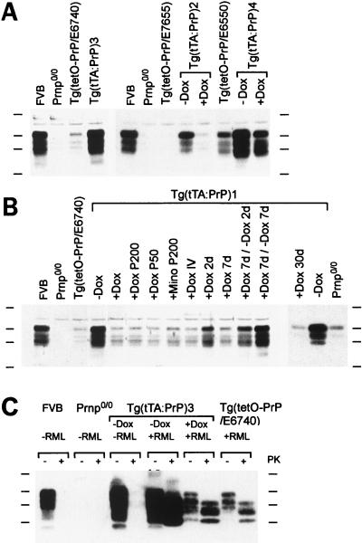
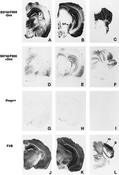
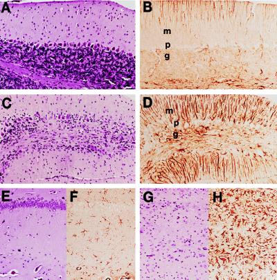
Conversion of the cellular prion protein (PrPC) into the pathogenic isoform (PrPSc) is the fundamental event underlying transmission and pathogenesis of prion diseases. To control the expression of PrPC in transgenic (Tg) mice, we used a tetracycline controlled transactivator (tTA) driven by the PrP gene control elements and a tTA-responsive promoter linked to a PrP gene [Gossen, M. and Bujard, H. (1992) Proc. Natl. Acad. Sci. USA 89, 5547-5551]. Adult Tg mice showed no deleterious effects upon repression of PrPC expression (>90%) by oral doxycycline, but the mice developed progressive ataxia at approximately 50 days after inoculation with prions unless maintained on doxycycline. Although Tg mice on doxycycline accumulated low levels of PrPSc, they showed no neurologic dysfunction, indicating that low levels of PrPSc can be tolerated. Use of the tTA system to control PrP expression allowed production of Tg mice with high levels of PrP that otherwise cause many embryonic and neonatal deaths. Measurement of PrPSc clearance in Tg mice should be possible, facilitating the development of pharmacotherapeutics.


Figures

Figure 1
Figure 1
PrPC and PrPSc in the brains of Tg(tTA:PrP) mice. Four lines of Tg(tTA:PrP) mice (Tables 1–3) were tested for PrP expression by Western blot analysis by using α-PrP polyclonal RO73 antiserum. Levels of expression in FVB, Prnp0/0, and Tg(tetO-PrP) lines are also shown. (A and B) Experimental conditions were as follows: “− Dox” denotes no treatment; “+ Dox” corresponds to doxycycline administered for 7 days in the drinking water at 2 mg/ml unless otherwise specified. “P50” corresponds to a 50 mg pellet (21-day release) of doxycycline placed subcutaneously for 7 days, and “P200” is a 200 mg pellet (21-day release). “IV” corresponds to a daily intravenous dose of 25 mg/kg of doxycycline for 3 days, and “d” is the abbreviation used for day. Induction of PrPC expression was observed in Tg(tTA:PrP) mice. Repression was obtained upon administration of doxycycline or minocycline (Mino) by using various routes and could be reversed upon ceasing antibiotic treatment. (C) Tg(tTA:PrP)3 animals left untreated (− Dox) and inoculated with RML prions (+ RML) were sacrificed at the time they presented neurological deficits consistent with development of scrapie (69 days postinoculation). Tg(tTA:PrP)3 mice treated with doxycycline (+ Dox) 1 week before inoculation as well as uninoculated Tg(tTA:PrP)3 and inoculated Tg(tetO-PrP/E6740) mice remained clinically healthy and were sacrificed at 200 days postinoculation. Brain homogenate consisting of 40 μg (A and B) or 60 μg (C) of protein was loaded per lane. Proteinase K digestion (20 mg/ml) was performed for 60 min at 37°C. Protein molecular weight markers from top to bottom correspond to 48, 35, 28, and 19 kDa.
Figure 2
Figure 2
Distribution of PrPC expression in the brain of Tg(tTA:PrP)3 mice. Distribution of MoPrPC is revealed by histoblots by using α-PrP polyclonal RO73 antiserum on half coronal brain sections of Tg(tTA:PrP)3 mice (AC) untreated or (DF) treated with oral doxycycline (2 mg/ml) for 30 days. Control brain sections from untreated (GI) Prnp0/0 and (JL) FVB mice. Tg(tTA:PrP)3 mice express PrPC at high levels in the hippocampus, neocortex, entorhinal cortex, substantia nigra (sn), and thalamus, as well as cerebellar granular (g) and molecular (m) layers. Although doxycycline treatment repressed PrPC expression, low levels of residual expression were still detected in the hippocampus, neocortex, entorhinal cortex, and granular layer of the cerebellum.
Figure 3
Figure 3
Doxycycline prevented neuronal loss, vacuolation, and gliosis in Tg(tTA:PrP)3 mice inoculated with RML prions. (A, B, E, and F) Tg(tTA:PrP)3 mice treated with oral doxycycline (2 mg/ml) and sacrificed at 200 days postintracerebral inoculation with RML prions. (C, D, G, and H) Tg(tTA:PrP)3 mice untreated with doxycycline and sacrificed at 70 days postintracerebral inoculation with RML prions when they showed multiple signs of CNS dysfunction; the first sign of neurologic disease was ataxia at 50 days after inoculation. (A, C, E, and G) Hematoxylin and eosin-stained brain sections. (B, D, F, and H) α-GFAP-immunostained brain sections. (AD) Views of cerebellar molecular (m), Purkinje cell (p), and granular (g) layers. (EH) Views of the hippocampus CA1 pyramidal cell layer (aligned in the upper part of Insets) with underlying stratum radiatum. Focal loss of Purkinje cells and granule cells was observed in the cerebellum of the ill Tg(tTA:PrP)3 mice and was accompanied by low grade vacuolation. Reactive astrocytic gliosis was demonstrated by the α-GFAP immunostaining that revealed moderate to severe gliosis in the granular and molecular layers (D) (Bergmann’s gliosis). In the hippocampal CA1 region most neuronal cell bodies were lost, and low grade vacuolation was apparent together with severe astrocytic gliosis (H).

References

    1. Prusiner S B. Science. 1997;278:245–251. - PubMed
    1. Büeler H, Fischer M, Lang Y, Bluethmann H, Lipp H-P, DeArmond S J, Prusiner S B, Aguet M, Weissmann C. Nature (London) 1992;356:577–582. - PubMed
    1. Manson J C, Clarke A R, Hooper M L, Aitchison L, McConnell I, Hope J. Mol Neurobiol. 1994;8:121–127. - PubMed
    1. Collinge J, Whittington M A, Sidle K C, Smith C J, Palmer M S, Clarke A R, Jefferys J G R. Nature (London) 1994;370:295–297. - PubMed
    1. Whittington M A, Sidle K C L, Gowland I, Meads J, Hill A F, Palmer M S, Jefferys J G R, Collinge J. Nat Genet. 1995;9:197–201. - PubMed

Publication types