

Fibrinogen deficiency reduces vascular accumulation of apolipoprotein(a) and development of atherosclerosis in apolipoprotein(a) transgenic mice
- PMID: 9770530
- PMCID: PMC22875
- DOI: 10.1073/pnas.95.21.12591
Fibrinogen deficiency reduces vascular accumulation of apolipoprotein(a) and development of atherosclerosis in apolipoprotein(a) transgenic mice
Abstract
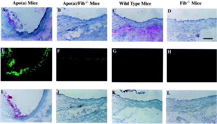
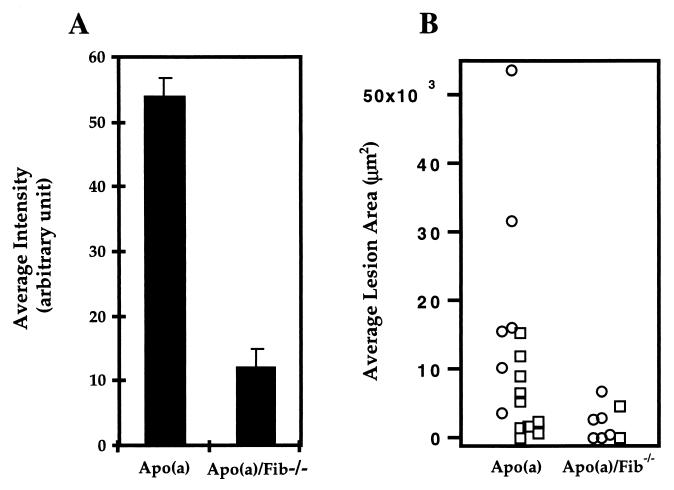
To test directly whether fibrin(ogen) is a key binding site for apolipoprotein(a) [apo(a)] in vessel walls, apo(a) transgenic mice and fibrinogen knockout mice were crossed to generate fibrin(ogen)-deficient apo(a) transgenic mice and control mice. In the vessel wall of apo(a) transgenic mice, fibrin(ogen) deposition was found to be essentially colocalized with focal apo(a) deposition and fatty-streak type atherosclerotic lesions. Fibrinogen deficiency in apo(a) transgenic mice decreased the average accumulation of apo(a) in vessel walls by 78% and the average lesion (fatty streak type) development by 81%. Fibrinogen deficiency in wild-type mice did not significantly reduce lesion development. Our results suggest that fibrin(ogen) provides one of the major sites to which apo(a) binds to the vessel wall and participates in the generation of atherosclerosis.
Figures
References
-
- Bostom A G, Cupples L A, Jenner J L, Ordovas J M, Seman L L, Wilson P W F, Schaefer E J, Castelli W P. J Am Med Assoc. 1996;276:544–548. - PubMed
-
- Assmann G, Schulte H, Eckardstein A V. Am J Cardiol. 1996;77:1179–1184. - PubMed
-
- Lawn R M, Wade D P, Hammer R E, Chiesa G, Verstuyft J G, Rubin E M. Nature (London) 1992;360:670–672. - PubMed
-
- Grainger D J, Kemp P R, Liu A C, Lawn R M, Metcalfe J C. Nature (London) 1994;370:460–462. - PubMed
-
- Edelberg J M, Gonzalez-Gronow M, Pizzo S V. Thromb Res. 1990;57:155–162. - PubMed
Publication types
MeSH terms
Substances
Grants and funding
LinkOut - more resources
Full Text Sources
Other Literature Sources
Molecular Biology Databases
