. 1998 Oct 13;95(21):12596-601.
doi: 10.1073/pnas.95.21.12596.

Down-regulation of transmembrane carbonic anhydrases in renal cell carcinoma cell lines by wild-type von Hippel-Lindau transgenes

Affiliations

Down-regulation of transmembrane carbonic anhydrases in renal cell carcinoma cell lines by wild-type von Hippel-Lindau transgenes

S V Ivanov et al. Proc Natl Acad Sci U S A. .

Abstract

To discover genes involved in von Hippel-Lindau (VHL)-mediated carcinogenesis, we used renal cell carcinoma cell lines stably transfected with wild-type VHL-expressing transgenes. Large-scale RNA differential display technology applied to these cell lines identified several differentially expressed genes, including an alpha carbonic anhydrase gene, termed CA12. The deduced protein sequence was classified as a one-pass transmembrane CA possessing an apparently intact catalytic domain in the extracellular CA module. Reintroduced wild-type VHL strongly inhibited the overexpression of the CA12 gene in the parental renal cell carcinoma cell lines. Similar results were obtained with CA9, encoding another transmembrane CA with an intact catalytic domain. Although both domains of the VHL protein contribute to regulation of CA12 expression, the elongin binding domain alone could effectively regulate CA9 expression. We mapped CA12 and CA9 loci to chromosome bands 15q22 and 17q21.2 respectively, regions prone to amplification in some human cancers. Additional experiments are needed to define the role of CA IX and CA XII enzymes in the regulation of pH in the extracellular microenvironment and its potential impact on cancer cell growth.


Figures

Figure 1
Figure 1
Schematic representation of the domain structure of wt and naturally occurring mut pVHLs. The three exons are indicated by different shadowing; the putative protein binding domains are represented by ovals, the filled one is the elongin binding domain; numbers above the pictures are amino acid residues, arrows indicate residues involved in producing mut pVHLs. Mut 1 is a 1-bp deletion creating a frameshift after residue 175; mut 2 is a 4-bp insertion creating a frameshift after residue 168; mut 3 and mut 4 are missense mutations, Gly-93–Asp and Tyr-98–His, respectively. Mut 1 and mut 2 are devoid of the elongin binding domain; mut 3 and mut 4 possess an intact functional elongin binding domain.
Figure 2
Figure 2
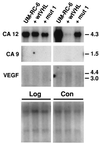
Northern analysis of target genes in growing (Log) and late confluent (Con) UM-RC-6 cells transfected with wt or mut 1 pVHL. Quantification of hybridization signals was done by using the PhosphorImager system (Molecular Dynamics). Before hybridization the blots were stained with methylene blue (Lower) to show approximately equal loading of the RNA samples (see also Materials and Methods). Numbers on the right represent size markers in kb.
Figure 3
Figure 3
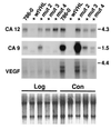
Northern analysis of target genes in growing (Log) and late confluent (Con) 786–0 cells transfected with wt or mut 2, mut 3, and mut 4 pVHL-containing plasmids. Quantification of hybridization signals was done by using the PhosphorImager system (Molecular Dynamics). Before hybridization the blots were stained with methylene blue (Lower) to show approximately equal loading of the RNA samples (see also Materials and Methods). Numbers on the right represent size markers in kb.
Figure 4
Figure 4
Global amino acid alignment of CA XII and CA IX proteins obtained by using the clustalw alignment tool as provided by the ExPasy server (http://expasy.hcuge.ch/sprot/scnpsite.html). The overall identity is 35.8%, and the identical residues are marked by ∗ below the sequence. The three essential Zn-liganded histidine residues are marked by Zn symbols above them. The short cytoplasmic domains are boxed with histidine, threonine, serine, and tyrosine residues underlined. The ideograms depicting the domain structure of the genes are positioned above the alignment. SP, signal peptide; CA, carbonic anhydrase domain; TM, transmembrane peptide; CTD, cytoplasmic domain; PG, proteoglycan domain.
Figure 5
Figure 5
Northern blot analysis of CA12 expression in normal tissues (A) and tumor cell lines (B) using the cDNA as hybridization probe. (A) The RNA filters are from CLONTECH (nos. 7759 and 7760) and contain 2 μg of poly(A)+ mRNA per tissue indicated: lanes 1, heart; 2, brain; 3, spleen; 4, thymus; 5, prostate; 6, testis; 7, ovary; 8, small intestine; 9, colon; 10, peripheral blood leucocytes. (B) The RNA filter is from CLONTECH (no. 775) and contains 2 μg of poly(A)+ mRNA per cell line indicated: lanes 1, promyelocytic leukemia, HL-60; 2, HeLa cells S3; 3, chronic myelogenous leukemia, K-562; 4, lymphoblastic leukemia, MOLT-4; 5, Burkitt’s lymphoma, Raji; 6, colorectal adenocarcinoma, SW 480; 7, lung adenocarcinoma, A549; 8, melanoma, G361, and poly(A)+ RNA from human nonsmall cell lung carcinomas (as indicated: 9, NCI H_1373; 10, NCI H_1264; 11, NCI H_1693; 12, NCI H_1944; 13, NCI H_838; 14, NCI H_1299; 15, NCI H_157; 16, NCI H_1466; 17, NCI H_460; 18, NCI H_727).
Figure 6
Figure 6
Subchromosomal localization of the human CA9 and CA12 genes. Metaphase after fluorescence in situ hybridization showing location of the (A) CA9 gene on the long arm of chromosome 17q21.2–21.3 (arrow) and (B) CA12 gene on the long arm of chromosome 15q22 (arrow). (Insets) Position of both loci, CA9 and CA12, on 4′,6-diamidino-2-phenylindole (DAPI)-banded human chromosomes, 17q21.2–21.3 and 15q22, respectively (arrows).

Similar articles

Cited by

References

    1. Latif F, Tory K, Gnarra J, Yao M, Duh F M, Orcutt M L, Stackhouse T, Kuzmin I, Modi W, Geil L, et al. Science. 1993;260:1317–1320. - PubMed
    1. Kishida T, Stackhouse T M, Chen F, Lerman M I, Zbar B. Cancer Res. 1995;55:4544–4548. - PubMed
    1. Duan D R, Pause A, Burgess W H, Aso T, Chen D Y, Garrett K P, Conaway R C, Conaway J W, Linehan W M, Klausner R D. Science. 1995;269:1402–1406. - PubMed
    1. Kibel A, Iliopoulos O, DeCaprio J A, Kaelin W G., Jr Science. 1995;269:1444–1446. - PubMed
    1. Duan D R, Humphrey J S, Chen D Y, Weng Y, Sukegawa J, Lee S, Gnarra J R, Linehan W M, Klausner R D. Proc Natl Acad Sci USA. 1995;92:6459–6463. - PMC - PubMed

Publication types

MeSH terms

Associated data