. 1998 Oct 13;95(21):12614-8.
doi: 10.1073/pnas.95.21.12614.

Deficient DNA-ligase activity in the metabolic disease tyrosinemia type I

Affiliations

Deficient DNA-ligase activity in the metabolic disease tyrosinemia type I

M J Prieto-Alamo et al. Proc Natl Acad Sci U S A. .

Abstract

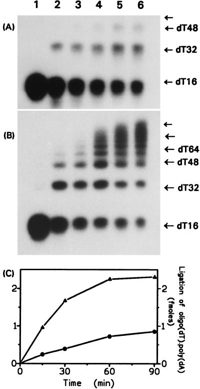
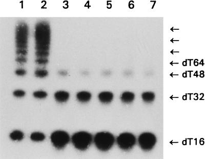
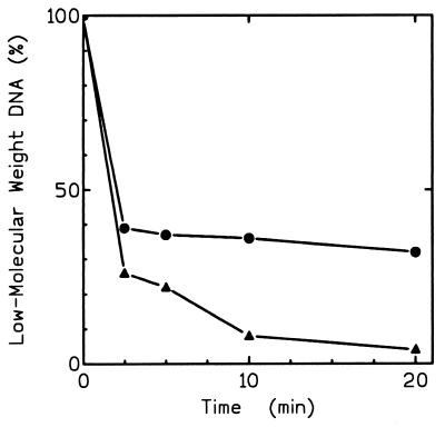
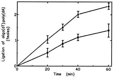
Hereditary tyrosinemia type I (HT1) is an autosomal recessive inborn error of metabolism caused by the deficiency of fumarylacetoacetate hydrolase, the last enzyme in the tyrosine catabolism pathway. This defect results in accumulation of succinylacetone (SA) that reacts with amino acids and proteins to form stable adducts via Schiff base formation, lysine being the most reactive amino acid. HT1 patients surviving beyond infancy are at considerable risk for the development of hepatocellular carcinoma, and a high level of chromosomal breakage is observed in HT1 cells, suggesting a defect in the processing of DNA. In this paper we show that the overall DNA-ligase activity is low in HT1 cells (about 20% of the normal value) and that Okazaki fragments are rejoined at a reduced rate compared with normal fibroblasts. No mutation was found by sequencing the ligase I cDNA from HT1 cells, and the level of expression of the ligase I mRNA was similar in normal and HT1 fibroblasts, suggesting the presence of a ligase inhibitor. SA was shown to inhibit in vitro the overall DNA-ligase activity present in normal cell extracts. The activity of purified T4 DNA-ligase, whose active site is also a lysine residue, was inhibited by SA in a dose-dependent manner. These results suggest that accumulation of SA reduces the overall ligase activity in HT1 cells and indicate that metabolism errors may play a role in regulating enzymatic activities involved in DNA replication and repair.


Figures

Figure 1
Figure 1
DNA-joining activity in ALV fibroblasts. Extracts (25 μg of proteins) from ALV (A) or normal (B) fibroblasts were incubated with the polynucleotide substrate [5′-32P]oligo(dT)16⋅poly(dA) at 37°C for 0 (lane 1), 15 (lane 2), 30 (lane 3), 60 (lane 4), 90 (lane 5), or 120 min (lane 6). The oligo(dT)16 multimers were separated in polyacrylamide/urea gels. The films were overexposed to detect the small amounts of multimers in A. (C) The data from A and B are expressed in femtomoles of oligo(dT)16 ligated. (•) ALV and (▴) normal fibroblasts.
Figure 2
Figure 2
DNA-ligase activity in fibroblasts from various HT1 patients. Extracts (25 μg of proteins) from normal fibroblasts (lane 1), LICH cells (lane 2), or fibroblasts from five unrelated HT1 patients (lanes 3–7) were incubated with the polynucleotide substrate [5′-32P]oligo(dT)16⋅poly(dA) for 120 min at 37°C. The oligo(dT)16 multimers were detected by electrophoresis in polyacrylamide/urea gels and autoradiography. For details, see Materials and Methods.
Figure 3
Figure 3
Joining of Okazaki fragments in normal and ALV fibroblasts. Cells synchronized in S phase were incubated in the presence of [α-32P]dATP for 1 min. The reaction then was chased by incubation with nonradioactive dATP for different times. After DNA isolation, replication intermediates were analyzed by gel electrophoresis and detected by autoradiography. The amount of low-molecular-weight DNA corresponding to Okazaki fragments was estimated by densitometry. (▴) Normal and (•) ALV fibroblasts.
Figure 4
Figure 4
T4 DNA-ligase activity in the presence of SA. T4 DNA-ligase was treated or not with increasing SA concentrations (15 min at 25°C) and then incubated at 37°C for 1 min with the oligo substrate. (A) The oligo(dT)16 multimers were separated in polyacrylamide/urea gels: T4 DNA-ligase without SA (lane 1) or incubated with increasing SA concentrations of 2.5(2), 5(3), 10(4), and 20(5) μM. (B) The activity was quantitated using an InstantImager (Packard). (C) Inhibition of enzyme-adenylate formation by SA. T4 DNA-ligase was incubated or not with increasing SA concentrations (15 min at 25°C) before the addition of [α-32P]ATP. The enzyme adenylate complexes were separated by electrophoresis and detected by autoradiography.
Figure 5
Figure 5
Influence of SA on the DNA-ligase activity present in normal cell extracts. Normal fibroblasts extracts (25 μg proteins) were preincubated for 15 min at 25°C without (▴) or with (•) SA (100 μM). The ligase activity was then measured as described in Materials and Methods.

Similar articles

Cited by

References

    1. Dehner L P, Snover D C, Sharp H L, Ascher N, Nakhleh R, Day D. Hum Pathol. 1989;20:149–158. - PubMed
    1. Lindblad B, Lindstedt S, Steen G. Proc Natl Acad Sci USA. 1997;74:4641–4645. - PMC - PubMed
    1. Tanguay R M, Valet J P, Lescault A, Duband J L, Laberge C, Lettre F, Plante M. Am J Hum Genet. 1990;47:308–316. - PMC - PubMed
    1. Phaneuf D, Labelle Y, Bérubé D, Arden K, Cavenee W, Gagné R, Tanguay R M. Am J Hum Genet. 1991;48:525–535. - PMC - PubMed
    1. Labelle Y, Phaneuf D, Leclerc B, Tanguay R M. Hum Mol Genet. 1993;2:941–946. - PubMed

Publication types