. 1998 Oct 13;95(21):12671-6.
doi: 10.1073/pnas.95.21.12671.

Galanin regulates prolactin release and lactotroph proliferation

Affiliations

Galanin regulates prolactin release and lactotroph proliferation

D Wynick et al. Proc Natl Acad Sci U S A. .

Abstract

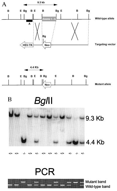
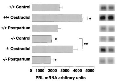
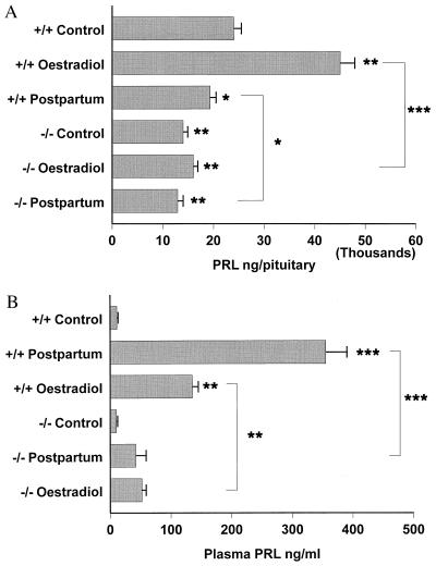
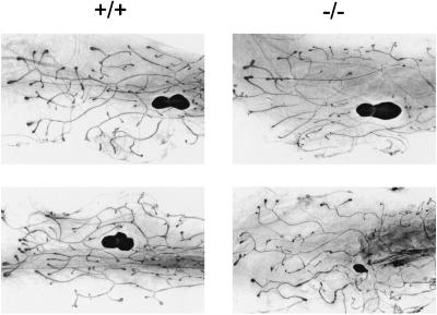
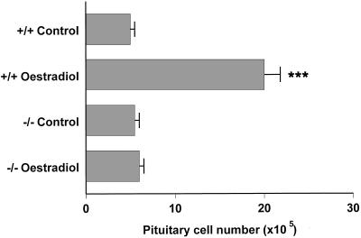
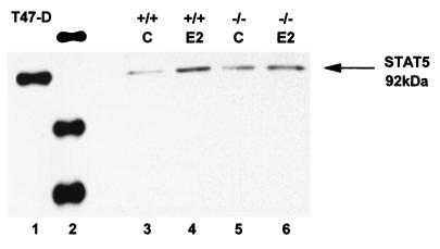
The neuropeptide galanin is predominantly expressed by the lactotrophs (the prolactin secreting cell type) in the rodent anterior pituitary and in the median eminence and paraventricular nucleus of the hypothalamus. Prolactin and galanin colocalize in the same secretory granule, the expression of both proteins is extremely sensitive to the estrogen status of the animal. The administration of estradiol-17beta induces pituitary hyperplasia followed by adenoma formation and causes a 3,000-fold increase in the galanin mRNA content of the lactotroph. To further study the role of galanin in prolactin release and lactotroph growth we now report the generation of mice carrying a loss-of-function mutation of the endogenous galanin gene. There is no evidence of embryonic lethality and the mutant mice grow normally. The specific endocrine abnormalities identified to date, relate to the expression of prolactin. Pituitary prolactin message levels and protein content of adult female mutant mice are reduced by 30-40% compared with wild-type controls. Mutant females fail to lactate and pups die of starvation/dehydration unless fostered onto wild-type mothers. Prolactin secretion in mutant females is markedly reduced at 7 days postpartum compared with wild-type controls with an associated failure in mammary gland maturation. There is an almost complete abrogation of the proliferative response of the lactotroph to high doses of estrogen, with a failure to up-regulate prolactin release, STAT5 expression or to increase pituitary cell number. These data further support the hypothesis that galanin acts as a paracrine regulator of prolactin expression and as a growth factor to the lactotroph.


Figures

Figure 1
Figure 1
(A) Targeted disruption of the murine galanin gene. The targeting vector replaces the first five exons of the galanin gene with a Neo cassette in reverse orientation. HSV-TK denotes the herpes simplex thymidine kinase and Neo the neomycin resistance gene. A denotes the 5′ external probe. B, BamHI, Bg, BglII, E, EcoRI., (B) Identical results obtained by Southern hybridization (genomic DNA digested with BglII and probed with the 5′ external probe) and PCR screening on the same litter derived from a mating of two heterozygotes.
Figure 2
Figure 2
Pituitary prolactin message levels in arbitrary units, measured by Northern blotting of randomly cycling wild-type and mutant females (denoted control), a second group of randomly cycling wild-type and mutant females treated for 3 weeks with estradiol-17β treatment (estradiol) and thirdly, another group of wild-type and mutant females 7 days postpartum. Values were determined relative to glyceraldehyde-3-phosphate dehydrogenase as a control probe. For all groups, n = 5. ∗, P < 0.05 denote levels of significance compared with the wild-type control group. Two representative pituitary samples are illustrated for each group.
Figure 3
Figure 3
Pituitary prolactin content in ng/pituitary (A) and circulating plasma prolactin in ng/ml (B) of randomly cycling wild-type and mutant females (denoted control), a second group of randomly cycling wild-type and mutant females treated for 3 weeks with estradiol-17β treatment (estradiol), and thirdly, another group of wild-type and mutant females seven days postpartum. n = 10 for all groups. ∗, P < 0.05, ∗∗, P < 0.01, ∗∗∗, P < 0.001 denote levels of significance compared with the wild-type control group.
Figure 4
Figure 4
Carmine-stained whole mounts of the fourth inguinal mammary gland from 8-week-old virgin wild-type (Left) and mutant (Right) animals. Two representative glands are shown for each genotype. Formation of the ductal tree is in progress, with mitotic terminal end buds clearly visible at the ductal termini; there is reduced ductal branching in the mutants animals. The lymph node is seen as a darkly staining mass at the center of the photographs.
Figure 5
Figure 5
Dispersed pituitary cell number in randomly cycling wild-type and mutant females (control) and after 3 weeks of estradiol-17β treatment (estradiol). For all groups, n = 10. ∗∗, P < 0.01, ∗∗∗, P < 0.001 denotes levels of significance compared with wild-type controls.
Figure 6
Figure 6
Representative Western blot of activated STAT5: lane 1, positive control using lysate of T47-D breast cancer cell line; lane 2, size marker; lanes 3 and 4, wild-type anterior pituitary lysates with and without estradiol-17β (E2); lanes 5 and 6, mutant anterior pituitary lysates with and without estradiol-17β; n = 5.

Similar articles

Cited by

References

    1. Goluboff L G, Ezrin C. J Clin Endocrinol Metab. 1969;29:1533–1538. - PubMed
    1. Orgnero-de-Gaisan E M, Maldonado C A, Aoki A. Histochem J. 1993;25:150–165. - PubMed
    1. Haggi E S, Torres A I, Maldonado C A, Aoki A. J Endocrinol. 1986;111:367–373. - PubMed
    1. Aoki A, de Gaisan E O, Pasolli H A, Torres A I. Exp Clin Endocrinol Diabetes. 1996;104:256–262. - PubMed
    1. Bakay L. J Neurosurg. 1950;7:240–255. - PubMed

Publication types

LinkOut - more resources