. 1998 Jul;43(1):56-63.
doi: 10.1136/gut.43.1.56.

Expression of inducible nitric oxide synthase activity in human colon epithelial cells: modulation by T lymphocyte derived cytokines

Affiliations

Expression of inducible nitric oxide synthase activity in human colon epithelial cells: modulation by T lymphocyte derived cytokines

G Kolios et al. Gut. 1998 Jul.

Abstract

Background: Nitric oxide (NO) synthesis and inducible nitric oxide synthase (iNOS) expression are increased in colonic biopsy specimens from patients with ulcerative colitis, but the cellular source of NO production is not known.

Aims: To examine the distribution of iNOS in human colonic mucosa and to explore the ability of T lymphocyte derived cytokines to regulate iNOS expression and activity in human colonic epithelial cells.

Methods: iNOS expression was examined using immunohistochemistry in colonic biopsy samples from 12 patients with ulcerative colitis and three with infectious colitis and compared with 10 normal controls. In vitro iNOS expression and activity were determined in HT-29 cell cultures; nitrite levels were measured using a fluorescent substrate, iNOS mRNA expression by northern blot analysis, and iNOS protein expression by western blot analysis.

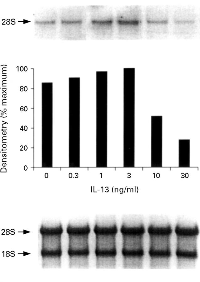
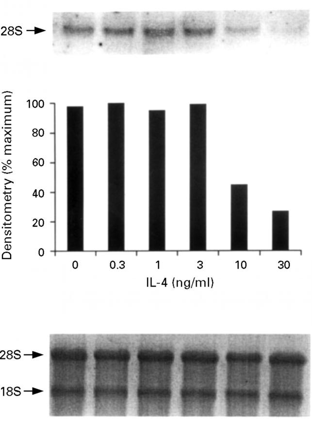
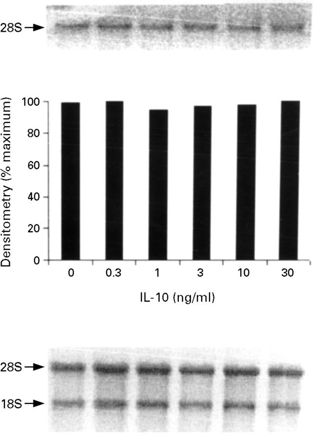
Results: No iNOS expression was detected (10 of 10) in non-inflamed mucosa derived from normal controls. In 11 of 12 cases of newly diagnosed ulcerative colitis, iNOS protein was expressed in the epithelial cells, while no other positive cells were found in the lamina propria. Similar iNOS labelling was found in colonic biopsy samples from patients with infectious colitis in the acute phase, but when re-examined in samples from patients in total remission, no iNOS staining was observed. Both interleukin (IL)-13 and IL-4, but not IL-10, are potent inhibitors of iNOS expression and activity induced by an optimal combination of cytokines, namely IL-1 alpha, tumour necrosis factor alpha and interferon gamma.

Conclusions: The data suggest that the epithelium is the major source of iNOS activity in ulcerative colitis and that IL-13 and IL-4 may act as intrinsic regulators of NO generation in intestinal inflammation.


Figures

Figure 1
Figure 1
All sections were stained with anti-human inducible nitric oxide synthase (iNOS) antibody (NO-53) using avidin/biotin peroxidase. (A) Normal bowel with no detection of iNOS. Original magnification × 100. (B) Ulcerative colitis (UC) with staining of superficial section of crypt and surface epithelium. Original magnification × 100. (C) Another UC case showing similar staining; iNOS labelling is located at the apical part of the epithelial cells, in close association with neutrophil infiltration of the lamina propria (arrowheads). Original magnification × 100. (D) Acute infectious colitis with staining of the superficial section of crypt and surface epithelium. Original magnification × 100. (E) Sample from the same patient after recovery, with resolution of iNOS expression. Original magnification × 50. (F) Same section as (D) using primary antibody preabsorbed with immunogenic peptide. Original magnification × 250.
Figure 2
Figure 2
Nitrite production by HT-29 cells after treatment with cytokines. Confluent monolayers of HT-29 cells were treated with interleukin (IL)-13 (30 ng/ml), IL-4 (30 ng/ml), IL-10 (30 ng/ml), IL-1α (10 ng/ml)/interferon γ (IFN-γ) (300 U/ml) or IL-1α (10 ng/ml)/IFN-γ (300 U/ml)/tumour necrosis factor α (TNF-α) (100 ng/ml), and after 48 hours incubation at 37°C nitrite levels were determined in supernatants, using a fluorescent substrate with a 10 nM level of detection. Basal is the amount of nitrite produced by HT-29 cells in the absence of added cytokines. Results are expressed as means (SEM) from three separate experiments.
Figure 3
Figure 3
Nitrite production by HT-29 cells after treatment with interleukin (IL)-1α (10 ng/ml)/interferon γ (IFN-γ) (300 U/ml)/tumour necrosis factor (TNF-α) (100 ng/ml) in the presence of increasing concentrations of IL-13. Proinflammatory cytokines were added after one hour of pretreatment with IL-13, and nitrite levels were determined in supernatants after 24 hours incubation at 37°C. Each point represents the mean (SEM) from three separate experiments. **p<0.01, ***p<0.001 compared with positive control response (no IL-13).
Figure 4
Figure 4
Nitrite production by HT-29 cells after treatment with interleukin (IL)-1α (10 ng/ml)/interferon γ (IFN-γ) (300 U/ml)/tumour necrosis factor α (TNF-α) (100 ng/ml) in the presence of increasing concentrations of IL-4. Proinflammatory cytokines were added after one hour of pretreatment with IL-4, and nitrite levels were determined in supernatants after 24 hours incubation at 37°C. Each point represents the mean (SEM) from three separate experiments. **p<0.01, ***p<0.001 compared with positive control response (no IL-4).
Figure 5
Figure 5
Inducible nitric oxide synthase (iNOS) mRNA expression by HT-29 cells after treatment with interleukin (IL)-1α (10 ng/ml)/interferon γ (IFN-γ) (300 U/ml)/tumour necrosis factor α (TNF-α) (100 ng/ml) in the presence of increasing concentrations of IL-13. The proinflammatory cytokines were added after one hour of pretreatment with IL-13 and incubated for 12 hours at 37°C. The top panel is the northern blot, the middle panel is the densitometry analysis of the blot, and the bottom panel the ethidium bromide stained 18S and 28S bands indicating equal loading of the lanes. This is a representative of three experiments.
Figure 6
Figure 6
Inducible nitric oxide synthase (iNOS) mRNA expression by HT-29 cells after treatment with interleukin (IL)-1α (10 ng/ml)/interferon γ (IFN-γ) (300 U/ml)/tumour necrosis factor (TNF-α) (100 ng/ml) in the presence of increasing concentrations of IL-4. The proinflammatory cytokines were added after one hour of pretreatment with IL-4 and incubated for 12 hours at 37°C. The top panel is the northern blot, the middle panel is the densitometry analysis of the blot, and the bottom panel the ethidium bromide stained 18S and 28S bands indicating equal loading of the lanes. This is a representative of three experiments.
Figure 7
Figure 7
Inducible nitric oxide synthase (iNOS) mRNA expression by HT-29 cells after treatment with interleukin (IL)-1α (10 ng/ml)/interferon γ (IFN-γ) (300 U/ml)/tumour necrosis factor α (TNF-α) (100 ng/ml) in the presence of increasing concentrations of IL-10. The proinflammatory cytokines were added after one hour of pretreatment with IL-10 and incubated for 12 hours at 37°C. The top panel is the northern blot, the middle panel is the densitometry analysis of the blot, and the bottom panel the ethidium bromide stained 18S and 28S bands indicating equal loading of the lanes. This is a representative of three experiments.
Figure 8
Figure 8
Inducible nitric oxide synthase (iNOS) protein expression by HT-29 cells after stimulation with interleukin (IL)-1α (10 ng/ml)/interferon γ (IFN-γ) (300 U/ml)/tumour necrosis factor α (TNF-α) (100 ng/ml) in the presence of increasing concentrations of IL-13. HT-29 cells were pretreated for one hour with IL-13, then proinflammatory cytokines were added. HT-29 monolayers were scraped after 24 hours incubation at 37°C and total protein was extracted. iNOS protein expression was determined by western blot analysis, using an anti-human iNOS antibody. This is a representative of three experiments.

Similar articles

Cited by

References

    1. Clin Exp Immunol. 1995 Jun;100(3):452-6 - PubMed
    1. Clin Exp Immunol. 1995 Sep;101(3):428-35 - PubMed
    1. Biochem J. 1996 Jan 15;313 ( Pt 2):641-6 - PubMed
    1. Eur J Immunol. 1996 Apr;26(4):934-8 - PubMed
    1. J Pediatr Gastroenterol Nutr. 1993 Apr;16(3):239-40 - PubMed

Publication types

MeSH terms