Comparative Study
. 1998 Jul;43(1):128-33.
doi: 10.1136/gut.43.1.128.

Periacinar stellate shaped cells in rat pancreas: identification, isolation, and culture

Affiliations
Comparative Study

Periacinar stellate shaped cells in rat pancreas: identification, isolation, and culture

M V Apte et al. Gut. 1998 Jul.

Abstract

Background: The pathogenesis of pancreatic fibrosis is unknown. In the liver, stellate cells (vitamin A storing cells) play a significant role in the development of fibrosis.

Aims: To determine whether cells resembling hepatic stellate cells are present in rat pancreas, and if so, to compare their number with the number of stellate cells in the liver, and isolate and culture these cells from rat pancreas.

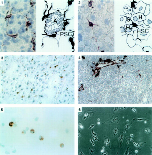
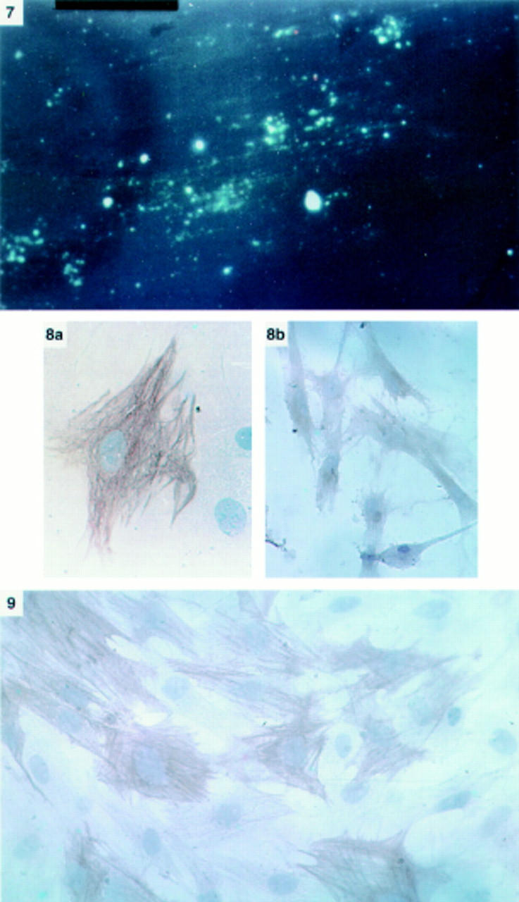
Methods: Liver and pancreatic sections from chow fed rats were immunostained for desmin, glial fibrillary acidic protein (GFAP), and alpha smooth muscle actin (alpha-SMA). Pancreatic stellate shaped cells were isolated using a Nycodenz gradient, cultured on plastic, and examined by phase contrast and fluorescence microscopy, and by immunostaining for desmin, GFAP, and alpha-SMA.

Results: In both liver and pancreatic sections, stellate shaped cells were observed; these were positive for desmin and GFAP and negative for alpha-SMA. Pancreatic stellate shaped cells had a periacinar distribution. They comprised 3.99% of all pancreatic cells; hepatic stellate cells comprised 7.94% of all hepatic cells. The stellate shaped cells from rat pancreas grew readily in culture. Cells cultured for 24 hours had an angular appearance, contained lipid droplets manifesting positive vitamin A autofluorescence, and stained positively for desmin but negatively for alpha-SMA. At 48 hours, cells were positive for alpha-SMA.

Conclusions: Cells resembling hepatic stellate cells are present in rat pancreas in a number comparable with that of stellate cells in the liver. These stellate shaped pancreatic cells can be isolated and cultured in vitro.


Figures

Figure 1
Figure 1
Panel 1: Immunostaining for desmin in pancreatic tissue sections (×40 objective). On the left is a photomicrograph of a section of the pancreas with a corresponding line diagram on the right. Desmin positive stellate shaped cells (PSC) were observed in the interstitium between acini (A). The cells had elongated cytoplasmic processes extending around the basal aspect of adjacent acinar cells. Panel 2: Immunostaining for desmin in liver tissue sections (×40 objective). On the left is a photomicrograph of a section of the liver with a corresponding line diagram on the right. Stellate cells (HSC) were stained brown and were located in the perisinusoidal space between hepatocytes (H) and hepatic sinusoids (S). Panel 3: Immunostaining for GFAP in pancreatic tissue sections (×25 objective). The positively stained (brown) stellate cells are seen in the same distribution as observed with the stain for desmin. Panel 4: Immunostaining for α-SMA in pancreatic tissue sections (×16 objective). No positively staining cells were found in the parenchyma of normal pancreas. Dense staining was observed in smooth muscle components of pancreatic ducts and blood vessels. Panel 5: Immunostaining for desmin in a cytospin of freshly isolated cells (×40 objective). Positive staining (brown) for desmin was observed in the cytoplasm. Panel 6: Phase contrast microscopy of cells after 24 hours in culture (×20 objective). Cells had assumed a flattened, angular appearance and showed abundant lipid droplets in the cytoplasm.
Figure 2
Figure 2
Panel 7: Vitamin A autofluorescence of cells after 24 hours in culture (×20 objective). Cells examined under ultraviolet illumination at 328 nm revealed blue-green fluorescence indicating the presence of vitamin A in the cytoplasm. Panel 8: Immunostaining for desmin (a) and GFAP (b) of pancreatic stellate shaped cells after 24 hours in culture (×40 and ×25 objective respectively). Cells were positive for both cytoskeletal markers. The high power view of the desmin positive cell clearly reveals a typical fibrillar staining pattern in the cell cytoplasm. Panel 9: Immunostaining for α-SMA of pancreatic stellate shaped cells after 48 hours in culture (×25 objective). Positive staining of cells was observed in a fibrillar pattern in the cytoplasm.

References

    1. Anat Rec. 1979 Aug;194(4):571-86 - PubMed
    1. Biol Cell. 1996;87(1-2):65-73 - PubMed
    1. Hepatology. 1984 May-Jun;4(3):392-403 - PubMed
    1. Hepatology. 1984 Jul-Aug;4(4):709-14 - PubMed
    1. Hepatology. 1987 Jul-Aug;7(4):680-7 - PubMed

Publication types