. 1998 Jul;43(1):134-9.
doi: 10.1136/gut.43.1.134.

Enhanced fibroblast growth factor 5 expression in stromal and exocrine elements of the pancreas in chronic pancreatitis

Affiliations

Enhanced fibroblast growth factor 5 expression in stromal and exocrine elements of the pancreas in chronic pancreatitis

T Ishiwata et al. Gut. 1998 Jul.

Abstract

Background: Fibroblast growth factor 5 (FGF-5) belongs to a group of mitogenic and angiogenic heparin binding growth factors but its potential role in chronic inflammatory conditions is not known.

Aims: To compare FGF-5 expression in the normal pancreas and in the pancreas of patients with chronic pancreatitis (CP) and to characterise FGF-5 expression and secretion in TAKA-1 cells, an immortalised Syrian hamster pancreatic duct cell line.

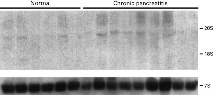
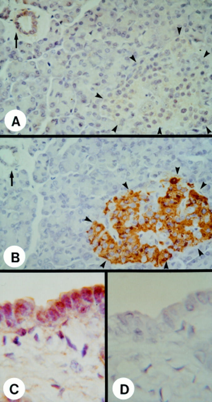
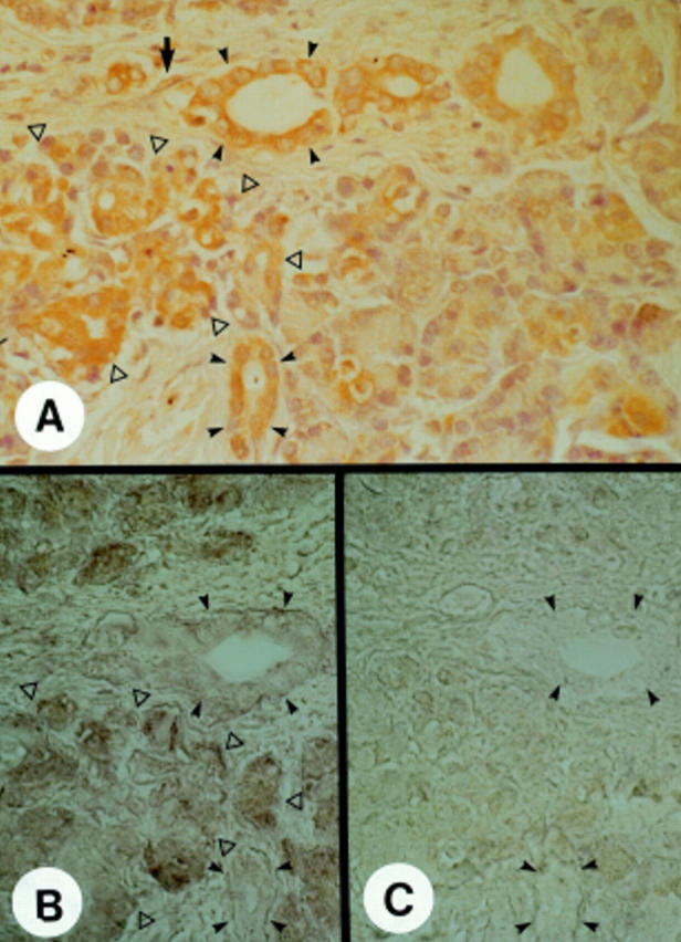
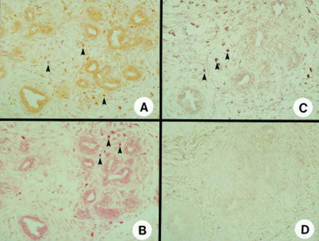
Methods and results: Northern blotting revealed the presence of a 4.0 kb FGF-5 mRNA transcript in both normal and CP tissue samples. Densitometric analysis indicated that the transcript levels were increased by a factor of 1.44 in CP tissue samples compared with normal tissue samples (p = 0.039). By immunohistochemisty and in situ hybridisation, FGF-5 was faintly expressed in ductal and islet cells in the normal pancreas. In contrast, in CP tissue samples, there was abundant expression of FGF-5 in ductal, acinar, and islet cells, as well as in periductal fibroblasts. FGF-5 was also expressed in TAKA-1 cells as determined by Northern blotting. By immunoblotting of heparinsepharose precipitates, TAKA-1 cells were shown to secrete FGF-5 into the medium.

Conclusion: Exocrine and stromal derived FGF-5 has the potential to participate in autocrine and paracrine pathways that may contribute to the pathobiology of chronic pancreatitis.


Figures

Figure 1
Figure 1
Expression of FGF-5 mRNA in the normal human pancreas and in chronic pancreatitis. Northern blotting of total RNA (20 µg/lane) was carried out using a 306 bp human FGF-5 cDNA fragment, randomly labelled with -32P]dCTP (750 000 cpm/ml; 14 day exposure). A mouse 7S cDNA probe, cross reactive with human 7S, was used as a loading control (40 000 cpm/ml; six hour exposure). rRNA markers are shown on the right.
Figure 2
Figure 2
Immunohistochemistry of FGF-5 in the normal pancreas. (A) Faint FGF-5 immunoreactivity was present in many ductal cells forming the small ducts (arrow) and, to a lesser extent, in islet cells (arrowheads). (B) Localisation of endocrine islets (arrowheads) in a serial section using an antiporcine insulin antibody, cross reactive with human insulin. The ductal cells forming the small ducts (solid arrow) were devoid of insulin immunoreativity. (C) Moderate FGF-5 immunoreactivity was present in the ductal cells of larger ducts and in the fibroblasts within the surrounding stroma. (D) Negative control without primary antibody did not show any positive immunoreactivity. Original magnification: ×500 (A,B); ×1250 (C,D).
Figure 3
Figure 3
Localisation and expression of FGF-5 in chronic pancreatitis. (A) Immunostaining showed moderate to strong FGF-5 immunoreactivity in the cytoplasm of ductal cells (solid arrowheads), acinar cells (open arrowheads), and fibroblasts (arrow). (B) In situ hybridisation analysis of serial sections revealed a mild to moderate FGF-5 mRNA signal in ductal cells (solid arrowheads) and fibroblasts, and a moderate to strong FGF-5 mRNA signal in acinar cells (open arrowheads). A sense FGF-5 probe (C) did not show any signal. Original magnification: ×500.
Figure 4
Figure 4
Localisation and expression of FGF-5 in the chronic pancreatitis-like regions adjacent to the cancer cells in the pancreatic cancer samples. (A) Immunostaining showed moderate to strong FGF-5 immunoreactivity in the cytoplasm of proliferating ductal cells, fibroblasts, and infiltrating macrophages (arrowheads), but not in the abundant connective tissue stroma. (B) Immunostaining of serial sections with alkaline phosphatase labelled streptavidin and new fuchsin also showed moderate to strong FGF-5 immunoreactivity in proliferating ductal cells, fibroblasts, and infiltrating macrophages (arrowheads). (C) In situ hybridisation analysis of serial sections revealed a moderate FGF-5 mRNA signal in proliferating ductal cells and fibroblasts, and a strong FGF-5 mRNA signal in the macrophages (arrowheads). In contrast, a sense FGF-5 probe (D) did not show any signal. Original magnification: ×250.
Figure 5
Figure 5
FGF-5 mRNA expression and FGF-5 protein secretion in TAKA-1 Syrian hamster pancreatic duct cells. (A) Total RNA (20 µg/lane) from TAKA-1 cells (lane 1) and COLO-357 human pancreatic cancer cells (lane 2, positive control) was used for northern blot analysis with a human FGF-5 cDNA probe (750 000 cpm/ml, four day exposure). A mouse 7S cDNA probe was used as a loading control (40 000 cpm/ml; eight hour exposure). rRNA markers are shown on the right. (B) Western blotting of the heparin-sepharose precipitates from two day conditioned serum free medium of TAKA-1 cells (lane 1), serum free medium (lane2), and serum free medium with 500 ng FGF-5 (lane 3) was carried out with a specific antihuman FGF-5 antibody (0.8 µg/ml, 45 minute exposure). Molecular weight markers are shown on the right.

Similar articles

Cited by

References

    1. Oncogene. 1987;1(4):369-76 - PubMed
    1. Oncogene. 1997 Sep 18;15(12):1417-24 - PubMed
    1. World J Surg. 1990 Jan-Feb;14(1):83-7 - PubMed
    1. Mol Cell Biol. 1991 Apr;11(4):1840-5 - PubMed
    1. Oncogene. 1991 Nov;6(11):2137-44 - PubMed

Publication types

MeSH terms