

Detection of Lassa virus antinucleoprotein immunoglobulin G (IgG) and IgM antibodies by a simple recombinant immunoblot assay for field use
- PMID: 9774554
- PMCID: PMC105290
- DOI: 10.1128/JCM.36.11.3143-3148.1998
Detection of Lassa virus antinucleoprotein immunoglobulin G (IgG) and IgM antibodies by a simple recombinant immunoblot assay for field use
Abstract
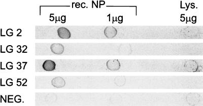
The nucleoprotein of Lassa virus, strain Josiah, was expressed in Escherichia coli as an N-terminally truncated, histidine-tagged recombinant protein. Following affinity purification the protein was completely denatured and spotted onto nitrocellulose membrane. A total of 1 microgram of protein was applied for detection of Lassa virus antibodies (LVA) in a simple immunoblot assay. Specific anti-Lassa immunoglobulin M (IgM) antibodies could be detected by increasing the amount of protein to 5 microgram. A panel of 913 serum specimens from regions in which Lassa virus was endemic and from regions in which Lassa virus was not endemic was used for evaluating the sensitivity and specificity of the LVA immunoblot in comparison to those of an indirect immunofluorescence (IIF) assay. The sera originated from field studies conducted in the Republic of Guinea (570 serum samples) and Liberia (99 serum samples), from inpatients of the clinical department of the Bernhard-Nocht-Institute, Hamburg, Germany (94 serum samples), and from healthy German blood donors (150 serum samples). In comparison to the IIF assay the LVA immunoblot assay had a specificity of 90.0 to 99.3%, depending on the origin of the specimens. The sensitivity was found to be highest for the Guinean samples (90.7%) and was lower for the Liberian samples (75%). Acute Lassa fever was diagnosed by PCR in 12 of 59 (20.3%) patients with fever of unknown origin (FUO) from the Republic of Guinea. On admission to the hospital, nine Lassa fever patients (75%) were reactive by the IgM immunoblot assay. One of the patients was infected with a new Lassa variant, which showed 10.4% variation on the amino acid level in comparison to the prototype strain of Lassa virus, Josiah. Seven PCR-negative patients were reactive by immunoblotting. The positive and negative predictive values of a single IgM immunoblot result for acute, PCR-confirmed Lassa fever were therefore 53.6 and 93.0%, respectively. Because of its high negative predictive value, a single IgM immunoblot result will be valuable for excluding acute Lassa fever for cases of FUO in areas where Lassa fever is endemic.
Figures

References
-
- Barber G N, Clegg J C S, Chamberlain J. Expression of Lassa virus nucleocapsid protein segments in bacteria: purification of high-level expression products and their application in antibody detection. Gene. 1987;56:137–144. - PubMed
-
- Barber G N, Clegg J C S, Lloyd G. Expression of the Lassa virus nucleocapsid protein in insect cells infected with a recombinant baculovirus: application to diagnostic assays for Lassa virus infection. J Gen Virol. 1990;71:19–28. - PubMed
-
- Clegg J C S, Lloyd G. Structural and cell-associated proteins of Lassa virus. J Gen Virol. 1983;64:1127–1136. - PubMed
-
- Clos J, Brandau S. pJC20 and pJC40—two high-copy-number vectors for T7 RNA polymerase-dependent expression of recombinant genes in E. coli. Protein Expr Purif. 1994;5:133–137. - PubMed
Publication types
MeSH terms
Substances
LinkOut - more resources
Full Text Sources
Other Literature Sources
