. 1998 Nov;36(11):3303-8.
doi: 10.1128/JCM.36.11.3303-3308.1998.

Molecular analysis of glycopeptide-resistant Enterococcus faecium isolates collected from Michigan hospitals over a 6-year period

Affiliations

Molecular analysis of glycopeptide-resistant Enterococcus faecium isolates collected from Michigan hospitals over a 6-year period

L Thal et al. J Clin Microbiol. 1998 Nov.

Abstract

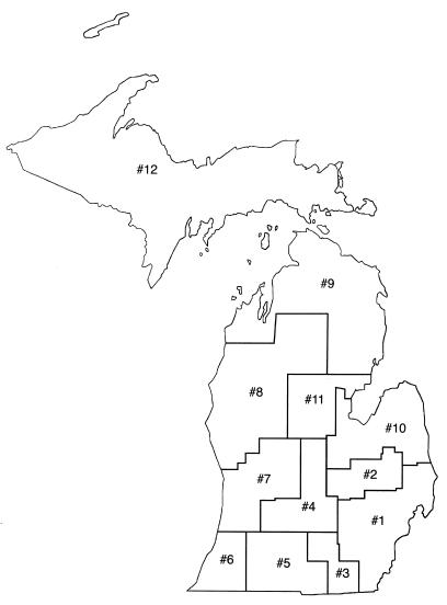
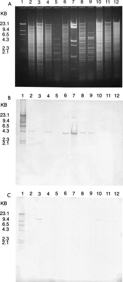
The purpose of this study was to evaluate the molecular relatedness of clinical isolates of glycopeptide-resistant Enterococcus faecium isolates collected from hospitals in Michigan. A total of 379 isolates used in this study were all vancomycin-resistant E. faecium isolates collected from 28 hospitals and three extended-care facilities over a 6-year period from 1991 to 1996. For the 379 isolates, there were 73 pulsed-field gel electrophoresis (PFGE) strain types. Within strain types, there were as many as six restriction fragment differences. Most isolates (70%) belonged to six strain types, which were designated M1 (36%), M2 (3%), M3 (18%), M4 (6%), M10 (4%), and M11 (3%). PFGE strain M1 was cultured from 135 patients in 13 hospitals during the period 1993 to 1996. Strain type M2 was cultured from 11 patients in two hospitals during the period 1991 to 1992 and was not observed after 1992. Strain type M3 was cultured from 70 patients in 10 hospitals during the period of 1994 to 1996. Both M4 and M10 were cultured from 23 patients in three hospitals and from 15 patients in two hospitals, respectively, during 1995 to 1996. M11 was cultured from 13 patients in four hospitals during 1996. A total of 23 of 28 hospitals had evidence of clonal dissemination of some isolates. Plasmid content and hybridization analysis done on 103 isolates from one hospital and two affiliated extended-care facilities indicated that the strains contained from one to eight plasmids. Mating experiments indicated transfer of vancomycin resistance from 94 of these isolates into plasmid-free E. faecium GE-1 at transfer frequencies of <10(-9) to 10(-4). Gentamicin resistance and erythromycin resistance were cotransferred at various frequencies. A probe for the vanA gene hybridized to the plasmids of 23 isolates and to the chromosomes of 72 isolates. A probe for the vanB gene hybridized to the chromosomes of 8 isolates. The results of this study suggest inter- and intrahospital dissemination of vancomycin-resistant E. faecium strains over a 6-year period in southeastern Michigan. The majority of isolates studied belonged to the same few PFGE strains, indicating that clonal dissemination was responsible for most of the spread of resistance that occurred.


Figures

FIG. 1
FIG. 1
Map showing 12 CHARs in Michigan.
FIG. 2
FIG. 2
PFGE of SmaI-digested genomic DNAs from VREF. Lanes: 1, lambda phage DNA ladder standard; 2, M1, most common strain type (36% of isolates) from CHAR 1; 3, M2 (3% of isolates) from CHAR 1; 4, M3 (18% of isolates) from CHARs 1, 4, and 11; 5, M4 (6% of isolates) from CHAR 1; 6, M10 (4% of isolates) from CHAR 1; 7, M11 (3% of isolates) from CHARs 1 and 4; 8, M20, from CHAR 8; 9, M22, from CHARs 1 and 8; 10, M23, from CHARs 1, 2, and 8; 11, M24 from CHAR 4; 12, M27 from CHAR 4; 13, M66 from CHAR 9; 14, M67 from CHAR 5; 15, M69 from CHAR 11; 4, 7, 9, and 10, strain types found in more than one CHAR; 1 to 6, 9, 10, and 12, strain types involved in interhospital dissemination; 8 and 11, strain types involved in intrahospital dissemination only; 13 to 15, unique strain types not involved in any dissemination.
FIG. 3
FIG. 3
(A) Agarose gel electrophoresis of EcoRI-digested genomic and plasmid DNAs from VREF isolates. Lanes: 1, biotin-labeled HindIII-digested lambda phage DNA; 2, chromosomal DNA from a known VanA strain; 3, chromosomal DNA from a known VanB strain; 4 and 5, genomic and plasmid DNA from a group 4 isolate (vanA gene on chromosome); 6 and 7, genomic and plasmid DNA from a group 4 isolate (vanA gene on plasmid); 8 and 9, genomic and plasmid DNA from a group 11 isolate (vanA gene on plasmid); 10 to 12, genomic DNAs from VanB isolates (group 23, group 8, and group 9, respectively). (B) Southern blot of gel shown in panel A probed with biotin-labeled vanA gene. Lanes: 1, biotin-labeled HindIII-digested lambda phage DNA; 2, chromosomal DNA from a known VanA strain positive for the vanA gene; 3, chromosomal DNA from a known VanB strain negative for the vanA gene; 4, genomic DNA from an isolate positive for the vanA gene; 5, plasmid DNA from isolate in lane 4 negative for the vanA gene; 6, genomic DNA from isolate positive for the vanA gene; 7, plasmid DNA from the isolate in lane 6 also positive for the vanA gene; 8, genomic DNA from isolate positive for the vanA gene; 9, plasmid DNA from the isolate in lane 8 positive for the vanA gene; 10 to 12, genomic DNAs from isolates negative for the vanA gene. (C) Southern blot of gel in shown in panel A probed with biotin-labeled vanB gene. Lanes: 1, biotin-labeled HindIII-digested lambda phage DNA; 2, chromosomal DNA from a known VanA strain negative for the vanB gene; 3, chromosomal DNA from a known VanB strain positive for the vanB gene; 4 to 9, genomic and plasmid DNAs from these isolates negative for the vanB gene; 10 to 12, genomic DNAs from these isolates positive for the vanB gene.
FIG. 4
FIG. 4
(A) CHEF electrophoresis of SmaI-digested genomic DNAs from VREF strains. Lanes: 1, lambda phage DNA ladder standard; 2, known VanA strain (vanA gene on the chromosome); 3 and 5, isolates from group 4 with the vanA gene on the chromosome; 4 and 6, isolates from group 4 with the vanA gene on a plasmid; 7 and 8, isolates from group 11 with the vanA gene on a plasmid; 9, isolate from group 3 with the vanA gene on a plasmid; 10, isolate from group 3 with the vanA gene on the chromosome. (B) Southern blot of gel in shown in panel A probed with biotin-labeled vanA gene and biotin-labeled lambda phage DNA. Lanes: 1, lambda phage DNA ladder standard; 2, known VanA isolate positive for the vanA gene; 3, 5, 7, 8, and 10, SmaI-digested genomic DNAs from these isolates positive for the vanA gene (in lanes 7 and 8, the vanA probe hybridizes with a plasmid visible on the CHEF gel); 4, 6, and 9, SmaI-digested genomic DNAs from these isolates negative for the vanA gene (plasmid DNA with the vanA gene is not visible on the CHEF gel).

Similar articles

Cited by

References

    1. Arthur M, Courvalin P. Genetics and mechanisms of glycopeptide resistance in enterococci. Antimicrob Agents Chemother. 1993;37:1563–1571. - PMC - PubMed
    1. Ausubel F M, et al., editors. Current protocols in molecular biology. 1, unit 2.4. New York, N.Y: John Wiley and Sons, Inc.; 1993.
    1. Bonten M, Slaughter S, Hayden M, Nathan C, Voorhis J V, Rice T, Weinstein R A. Abstracts of the 36th Interscience Conference on Antimicrobial Agents and Chemotherapy, 15 to 17 September 1996, New Orleans, La. Washington, D.C: The American Society for Microbiology; 1996. Patients’ endogenous flora as a source of “nosocomial” vancomycin-resistant enterococci (VRE)
    1. Boyce J M, Opal S M, Chow J W, Zervos M J, Potter-Bynoe G, Sherman C B, Romulo R L C, Fortna S, Medeiros A A. Outbreak of multidrug-resistant Enterococcus faecium with transferable vanB class vancomycin resistance. J Clin Microbiol. 1994;32:1148–1153. - PMC - PubMed
    1. Centers for Disease Control. Nosocomial enterococci resistant to vancomycin—United States, 1989–1993. Morbid Mortal Weekly Rep. 1993;42:597–599. - PubMed

Publication types

MeSH terms

LinkOut - more resources